What is calcific tendinitis?

Calcific tendinitis occurs when calcium deposits builds up in the tendons. Calcific tendinitis most often occurs in the shoulders. Calcific tendinitis can happen in elbows, wrists, hands, hips, knees or feet as well.

Calcific Tendonitis

What causes calcific tendinitis?

Calcific tendinitis is a process whereby calcium deposits are buildup and cause a chemical reaction with other tissues in the tendon to cause pain. In the shoulder, the calcium deposits is usually accumulated inside the rotator cuff.

What are the symptoms of a calcific tendinitis?

You may experience pain and stiffness that often come and goes and usually lasts for 1 to 2 months. You may find it difficult to sleep at night due to the pain.

How is calcific tendinitis diagnosed?

Your doctor will physically examine your affected area. An X-ray may ordered to look for the calcium deposits. Additional investigation like Ultrasound or MRI may be required to see if there are any tears to the tendons and other soft tissues.

How is calcific tendinitis treated?

If you are in extreme pain, you may want to seek medical attention.

Treatments at clinical level includes:

Book Appointment Online for Calcific Tendinitis Treatment

What is the collarbone?

The collarbone (clavicle) is one of the bones of the shoulder joint. It holds the shoulder, the shoulder blade and the sternum. The collarbone also protects the nerves and the blood vessels from the neck to the shoulder.

Broken Collarbone

What causes a broken collarbone?

A broken collarbone is cause by a direct impact injury to the collarbone or to the shoulder. People involve in sports such as football, wrestling or ice hockey are at more risk of getting broken collarbone.

The collarbone is a common bone that gets broken. Young person age between 13 to 20 break it most often. Young children have a high chance of getting a broken bone during play.

What are the symptoms of a broken collarbone?

Symptoms of a broken collarbone are:

  • Pain felt immediately after the fall or the collarbone was hit.
  • Unable to raise the arm of the injured site.
  • Grinding feeling felt when trying to raise the injured arm.

You will see deformity in some cases. If you see a deformity, you will probably seeing a bump or swell along the collarbone, the shape of the collarbone is different compare to the uninjured collarbone. The bone may push the skin out, causing a tent-shaped look.

How is a broken collarbone diagnosed?

A broken collarbone is usually detected by an X-ray. The X-ray can tell the severity of the break.

Fracture Clavicle

Your doctor will be looking:

  • For deformity at your collarbone.
  • How well is the range of motion of your shoulder.
  • The strength of your shoulder

How is Broken Collarbone Treated?

Surgery is recommended for patients with severe break to the collarbone or they prefer a faster healing process. If the bone is displaced, surgery is a more suitable option to fix the fracture. Many young and active people prefer surgery.

After surgery, you will use a sling for up to a week. Your doctor will teach you light exercises to move your shoulder. Usually you can resume sports after a month.
Book Appointment Online for Broken Collarbone Treatment

Osteoarthritis

If you think osteoarthritis is the disease of the old, you may be wrong. Osteoarthritis (OA) is a degenerative joint disease where the cartilage in the joints wears down over time. Any joint could be affected, but the majority of the OA occurs at the joints on the hands, knee, hips, lower spine or neck.
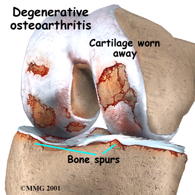
Osteoarthritis Knee

The most common OA symptoms include pain, tenderness at the joint area, stiffness in the morning or after resting for long hours and the feeling of creaking at the joints. These symptoms occur slowly and worsen with time. As the condition gets more severe, bone protrusion around the joint area may be formed.

Osteoarthritis of the Knee

Know what is Osteoarthritis

In most cases, the exact cause of the osteoarthritis is unknown. However, some risk factors have been identified to contribute to the wear and tear of the joint cartilage and lead to OA. Women have a higher risk of developing OA with an unknown reason. OA usually occurs in older adults, rarely in people under the age 40. For those younger individuals, genetic conditions where the joints or the cartilage is malformed could increase the likelihood of developing OA. Besides, any injuries to the joints due to sports or accidents may also cause OA, both in young and old people. Obesity is another risk factor in OA development as more stress is incurred on the weight bearing joint especially the knees.

Check whether you have Osteoarthritis

If you always suffer from pain in your joints, do have a check with your healthcare professionals. X-ray is the most common non-invasive method to determine the degree of cartilage loss in the joints and is also to rule out possibilities of other joint or bone problems. There is no blood test to diagnose OA but blood test results are important to reveal other diseases which could resemble symptoms of OA, for example rheumatoid arthritis (an auto-immune condition of the joints). Other tests include drawing fluid out from the joint using needle (a procedure called arthrocentesis) to exclude the possibilities of gout of infection as the cause of joint pain and swelling.

Treat Osteoarthritis Today

Non-surgical method to treat osteoarthritis:

  • Viscosupplementation (Hyaluronic Injection) – Lubricant injection to the joint.
  • Anti-inflammatories – Reduce inflammation to the joints.
  • Physiotherapy – Trains certain sets of muscles to build strength to the joints.

Surgical intervention to treat severe osteoarthritis:

Exercises to Prevent Osteoarthritis

Light exercise such as biking or swimming is often recommended as these exercises could strengthen the muscles around the joint. Exercise also helps to reduce weight, which could in turn relieve some pressure on the joints thus relieving pain.

Avoid any forms of exercise if your joint is injured or swollen. Always rest your joints whenever there is pain or doing any repetitive movements that involve the joint. Besides, the use of assistive devices such as walking sticks allows for normal movement while reducing stress on the joints.

Glucosamine Helps Osteoarthritis

The use of glucosamine supplements alone has yielded some promising results in scientific studies. More well-designed studies are required to prove conclusively the benefits of these health supplements on OA prevention or treatment. Nevertheless, do check with your healthcare professional before using any health supplements for OA.
Book Appointment Online for Osteoarthritis Treatment

Osteoporosis

Osteoporosis is a common condition in which the bones are brittle due to a low bone mass density or reduction in bone strength. As a result, a simple fall may actually fracture any brittle bones especially at the hips, wrist or the spine.

How soon could one be affected by Osteoporosis?

Osteoporosis is an age-related disease which is more common in women than men. Women with early menopause before the age 45, either naturally or through surgical removal of the ovaries, are at higher risk of developing osteoporosis. Besides, elderly who are confined in bed or with low physical activity may be more prone to osteoporosis. Men or some individuals may get osteoporosis at a younger age if they are on long term intake of some medication (e.g. steroids, thyroid medications) and certain medical conditions (e.g. rheumatoid arthritis, hyperparathyroidism, eating disorder) place a person at a higher risk. Smoking and excessive alcohol consumption have been greatly associated with the increased incidences of osteoporosis well.

What are the symptoms of Osteoporosis for men and women respectively?

Osteoporosis

Osteoporosis gradually develops over the years and may not have any signs or symptoms during the early stages. The symptoms usually appear in its advanced stages where fractures have occurred. Depending on the location of the fractured bone, one may feel pain at the affected area, for instance pain at the neck , lower back may indicate spinal bone fracture. Some may experience a loss of height over time or develop a stooped posture (also known as “dowager hump” as it is more common in women).  Nevertheless, symptoms of osteoporosis in men are similar to that in women.

Tips on daily Bone Care for women

Regular physical activities especially the weight bearing exercises (e.g. tai-chi, yoga, aerobics) and resistance exercise (e.g. weight-lifting) can help build stronger bone and maintain healthy bone density. It is important to choose the correct exercises for your age and existing health conditions to prevent unnecessary injury. Besides, calcium intake either from natural food sources (e.g. milk products, green leafy vegetables, bean curd) or supplementation of at least 1,000mg coupled with vitamin D is recommended especially for women of menopausal age. Quitting smoking and the reduction of alcohol consumption are both beneficial to prevent or slow down the progression of osteoporosis.

Osteoporosis Self-assessment Tool for Asians (OSTA) is a quick and easy tool to assess the risk of developing osteoporosis. The current available OSTA chart is developed for Asians aged 45 and above and by using just the present weight and age, one can determine their osteoporosis risk of either being too high, moderate or low.

Bone Mineral Density (BMD) screening is the best way to diagnose osteoporosis especially if your OSTA shoes that your are in high or moderate risk group. However, even if you are in the low risk group, BMD screening is still recommended if you have any risk factors mentioned earlier.
Book Appointment Online for Osteoporosis Treatment
OSTA

Arthroplasty is a surgical procedure done to reconstruct or replace a diseased joint. Arthroplasty for rheumatoid arthritis is done to correct the deformity or restore the function to the joint. Bones in a joint can be replaced with metal or plastic parts.

Knee Replacement X-ray

What To Expect After Arthroplasty for Rheumatoid Arthritis

Recovery after arthroplasty is gradual. You may need to stay in the hospital for 2 days and the next day after the surgery, you will be able to walk with the help of walking aids. Depending on the the joint operated, post operative rehabilitation program will take a few weeks.

Why Arthroplasty for Rheumatoid Arthritis Is Done

Arthroplasty for rheumatoid arthritis will provide pain relief and improve the range of movement of the joint to a certain extent.

Arthroplasty for rheumatoid arthritis is considered when:

  • Symptoms can no longer be controlled with medicine, joint injections, physical therapy, and exercise.
  • Pain from rheumatoid arthritis can no longer be tolerated.
  • You are not able to do normal daily activities.
  • Narrowing of the joint space or wearing away of the cartilage and bone is causing severe pain or reduced range of motion.

How Well Arthroplasty Works for Rheumatoid Arthritis

Arthroplasty can help in reducing the pain and restore  the function of the joint to allow you to resume to your normal activities.

What are the Risks of Arthrosplasty

The risks of arthroplasty include risks of a surgery and anaesthesia, the risks are small:

  • Infection
  • Loose joints

What To Think About For Arthroplasty For Rheumatoid Arthritis

Click to see more for the below surgery:

An important factor to the success of arthroplasty also depends on the person’s commitment to post operative rehabilitation.

Book Appointment Online for Arthroplasty Treatment