Improperly-fitting shoes, which can cause bunions, corns, calluses, hammertoes, and other disabling foot problems, are a huge public health risk. The following are statistics from the US, but are most likely applicable to Singapore, too, as a lot of women, for vanity’s sake are willing to endure punishment for their feet, in the name of fashion and beauty!

  • Nine out of 10 women are wearing shoes that are too small for their feet.
  • Eight out of 10 women say their shoes are painful.
  • More than seven out of 10 women have developed bunion, hammertoe, or other painful foot deformity.
  • Women are nine times more likely to develop a foot problem because of improper fitting shoes than a man.
  • Nine out of 10 women’s foot deformities can be attributed to tight shoes.

Bunions

The most common foot problems are bunions. Bunions are caused by wearing shoes that are too small. If the joint that connects your big toe to your foot has a swollen, sore bump, you may have a bunion.

Tailor's Bunion

More than half the women have bunions, a common deformity often blamed on wearing tight, narrow shoes, and high heels. If the painful swollen lump is on the outside of your foot near the base of your little toe, then it is called a bunionette (tailor’s bunion). An advanced bunion may make your foot look grotesque. If your bunion gets too severe, it may be difficult to walk. Your pain may become chronic and you may develop arthritis.

Arch Pain (Plantar Fasciitis)

Plantar fasciitis is one of the most common causes of heel pain. It is due to inflammation of a thick band of tissue called the plantar fascia, which is a fibrous band of tissue running from the heel to forefoot, along the bottom of the foot. Over or under pronation causes excessive shock to the foot when walking and running. The pain is localised in the heel and / or arch and is often extreme in the morning when first getting out of bed after prolonged rest. The chief diagnostic sign of this problem is pain at the bottom of the heel or arch when first standing, which gradually improves with walking. This pain may later return with continued walking. The pain usually subsides after a period of rest.

 Achilles Tendinitis
Achilles Tendinitis
Achilles Tendinitis

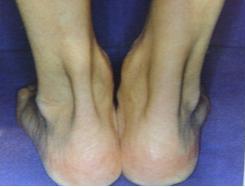
The Achilles tendinitis is a syndrome of irritation of the Achilles tendon in the ankle. The Achilles tendon is the large tendon in the back of the ankle that inserts into the heel bone. When individuals overuse their Achilles tendon, it becomes irritated and inflamed. This inflammation can cause pain and swelling and can lead to small tears within the tendon and make the Achilles tendon susceptible to rupture. This is often caused by repetitive, minor impact on the affected area, or from a sudden more serious injury. Incorrect posture at work or home or poor stretching or conditioning before exercise or playing sports also increases the risk.

Ingrown Toenails

Ingrown Toenail
Ingrown Toenail

An ingrown toenail occurs when a sharp corner of the toenail digs into the skin at the end or side of the toe. Pain and inflammation at the spot where the nail curls into the skin occurs first. The inflamed area then starts to grow extra tissue or drain yellowish fluid. Usually caused by tight-fitting shoes or high heels, the toes tend to get compressed together and pressurise the nail to grow abnormally. Improper trimming of toenails can also cause the corners of the nail to dig into the skin. Disorders such as fungal infections of the nail can also cause a thickened or widened toenail to develop.
Book Appointment Online for  Treatment

Osteoporosis  Screening Package @ $379

(Includes Specialist Consultation & BMD Screening).

Book Appointment Online for Osteoporosis Treatment

Drinking Milk is Not Enough to Prevent Osteoporosis!

Women, as they grow older, and especially after menopause, lose up to 20 percent of their bone mass in the five to seven years after menopause! Although osteoporosis affects men as well, in the last three decades since 1960s, the number of osteoporotic hip fractures in Singapore rose five-fold per 100k population in women over the age of 50.

In April, the Osteoporosis Society of Singapore [OSS] launched a campaign encouraging postmenopausal women to take early action to protect their bone health, instead of delaying to after their first bone fracture.

Singapore has the highest number of osteoporotic fractures in Asia yet many postmenopausal women, to the frustration of doctors here, are leaving it to after they have had their first fracture before taking preventive action.

Studies show that one in every five patients usually dies within the first year after a hip fracture. Of those who survive, more than half will lose their ability to walk and will end up bedridden or wheelchair-bound.

Drinking your calcium in the form of milk while good for bone health is not enough for those at risk of postmenopausal osteoporosis.

You Might be of High Fracture Risk due to Osteoporosis

Once you have menopaused, the OSS is encouraging postmenopausal women to undergo bone mineral density (BMD) screening.

The OSTA test will determine if a postmenopausal woman is at high, moderate or low risk of having a low BMD. Those who are at high risk or moderate risk with other risk factors should have their BMD tested.

OSTA
Patients with pre-existing osteoporotic fractures may heighten their risk of further fractures by two to four times. Therefore it will be prudent for patients with high risk of fractures to seek medical attention before their first fracture. The situation is expected to worsen when the proportion of Singapore population aged above 65 triples by 2030.

Too Late for More Milk to Prevent Osteoporosis

For osteoporotic patients, where the bone density has begun degenerating, medication is necessary as osteoporosis is a progressive one.

In a normal situation, drinking your milk and eating your cheese and sunning yourself for vitamin D will be good measures to build bone health but will not be sufficient when you identified with the bone degenerative condition – osteoporosis.

When faced with this, you need to medicate under your doctor’s supervision.

The International Osteoporosis Foundation reports that patients on medication reduce their risk for fracture by about 30-70%. But not all patients are found to be compliant.

Many stop taking their prescribed medication or fail to take it regularly. This is largely because osteoporosis is not associated with any visible to the eye symptoms. As a result, many fail to realise the hidden severity of osteoporosis until it is too late – after a fracture.

Patient compliance with prescribed treatment is most important whether they take oral medication or opt for the convenience of injections.

Patients who find it hard to comply with the regime of taking oral medication regularly, can consider other options such as yearly intravenous infusions.

Those with previous fractures, high-risk scores on OSTA should seek their doctor’s advice right away. Without medication, their risks of further fractures double or rise as high as four times.
Book Appointment Online for Osteoporosis Treatment

Managing Skin Sores

Skin sores are characterised by the appearance of blisters, bumps or ulcers on the skin around the body.

Some common causes of skin sores include:

  • Underlying health conditions (e.g. eczema, diabetic leg ulcers, skin cancer)
  • Pressure damage (e.g. skin sores)
  • Infection (e.g. cold sores, chicken pox)
  • Burn (e.g. scald, sunburn, frostbite, chemical burn, etc)
  • Injury (e.g. abrasion, cut)
  • Other situations (e.g. insect bites or stings, food allergies, medication side effects or allergies)

Managing Skin Sores:

Skin sores are skin damage as a result of prolonged pressure on the skin coupled with friction.

Due to the pressure, blood flow to the area is reduced, causing tissue damage and sores formation.

Skin sores are more common at areas where the bones are close to the skin (e.g. ankle, back, elbow, hips or buttocks). Bed or wheel-chair bound patients are at a higher risk of developing skin sores.

Skin sores are classified into 4 stages:

Stage 1

Intact skin with discolouration, the skin is firm, soft and with slight pain.

Stage 2

A superficial open wound that resembles a ruptured fluid-filled blister.

Stage 3

A deep wound with a crater-like appearance which may expose some fatty tissues or yellowish dead tissues.

Stage 4

Extensive loss of tissues which may expose the muscles or bones, with dark dead tissues at the bottom.

TIPS:

Stage 1 and 2 skin sores could be managed at home while the higher stages require medical attention as there is an increased risk of infection.

Preventing Diabetic Ulcers

  • Blood sugar levels should be well controlled in a diabetic patient.
  • Proper fitting footwear with adequate supper is important to minimise injury.
  • Minor foot injuries or foot infections (e.g. fungal) should be well-managed.
  • To maintain a clean and healthy foot, clean the foot gently with mild soap and water, pat dry and apply moisturisers.
  • Diabetic patients should have their feet checked regularly by an orthopaedic surgeon.

Self-management for skin sores and diabetic ulcers

A good wound dressing (e.g. foam dressings) for skin sores or diabetic leg ulcers should minimise pain and trauma during dressing change while keeping a moist wound environment. All these factors help to create an ideal environment with minimal wound disturbance for optimal wound healing.

You should see a doctor:

  • If the sore does not seemed to be healing.
  • If there is unbearable pain or the area becomes swollen with foul odour discharge.
  • If you are down with fever.
  • If you experience other new unexplainable symptoms (e.g. nausea)

Book Appointment Online for Skin Sore Treatment

Surgical Nail Removal Overview

You may want to consider surgical nail removal for severe or recurrent fungal nail infections. The whole nail or only a portion of the nail can be removed surgically.

Surgical nail removal can be done at the clinic. The orthopaedic surgeon will give an injection in the finger or toe for local anaesthesia. He will then start by loosen the skin around the nail and then tries to separate the nail from the skin by using a tool under the nail. If a portion of the nail is infected, only the affected nail is removed.

Our orthopaedic surgeon can remove the root of the nail to prevent the nail from growing back if you have recurrent nail infection that does not seems to go away with conservative treatment.

Your finger or toe will be covered with gauze and tape once the surgery is done.

What to Expect After the Surgical Nail Removal

For the first few weeks after the surgery, keep the wound dry and clean. The wound should heal very well after a few weeks.

Is it Necessary to Have My Nail Surgically Removed?

Surgical nail removal is performed when a large portion of the nail is infected and damaged or the nail is causing you extreme pain. Majority of the cases only requires to remove a portion of the affected nail.

How Well Does Surgical Nail Removal Works?

After the infected nail has been removed, the infection can be further treated by taking oral anti-fungal medicine.

What are the Risks Involved in Surgical Nail Removal?

The risk is very small. It could be:

  • Pain.
  • Infection.
  • Nail grows abnormally.

What to Think About for Surgical Nail Removal?

If you have been having recurrent nail infection, you can choose to have the nail removed together with its root. Once the root of the nail is removed, there is no chance for the infected nail to grow back again.
Book Appointment Online for Nail Removal Treatment

Relieves joint pain & improves mobility to put the spring back in your step and choices back into your life!

Synvisc (sodium hyaluronate) is a treatment to provide relief from the pain of osteoarthritis (OA). Now you have an alternative to the use of pain killers such as nonsteroidal anti-inflammatory drugs (NSAIDs) or steroids.

Given by your doctor, this proven treatment reduces joint pain and improves joint mobility allowing you to resume some of the activities you once enjoyed.

Synvisc Benefits: Lubricates, cushions and protects the joint

Synvisc renews more efficient lubrication and cushioning absent from the synovial fluid of an arthritic joint. It helps to restore essential properties of the synovial fluid.

Synvisc helps to lubricate, cushion and protect the joint, bringing relief that lasts up to 6 months, from one simple injection.

Synvisc is a simple procedure

The procedure is called visco-supplementation – a simple procedure done by an orthopaedic surgeon. Your doctor injects Synvisc directly into the joint where it combines with the synovial fluid.

For many patients, relief occurs after a few days and optimum results shortly, thereafter. Unlike other treatments that may require up to five injection, Synvisc only requires a single injection.

Synvisc – Pure and Simple. Helps you get back in the game!
Visit www.OrthopaedicSurgeon.com.sg

Important benefits of having Synvisc treatment

Patients can look forward to some of Synvisc advantages:

  • Significant pain reduction from osteoarthritis
  • Enhanced joint mobility and joint protection
  • Long term relief achieved with one single injection
  • Resumption of many activities you once enjoyed

Synvisc is made from a natural product

Synvisc is a highly purified, clear fluid made from a natural substance called sodium hyaluronate, which has been used in medicine for over 20 years. Synvisc has been specially formulated for single injection therapy. Your doctor can give you Synvisc by injection directly into the joint to relieve the pain of osteoarthritis at that site.

Synvisc added benefits of local treatment

  • Synvisc avoids the side effects often seen with traditional systemic osteoarthritis treatments
  • Synvisc injected into the joint at that site of discomfort restores the viscosity of the synovial fluid

Discuss Synvisc with your doctor

You and your doctor can decide if Synvisc is right for you. Ask for more information about the benefits of this pure and simple way to relieve osteoarthritis pain and enhance joint function.

How the knee joint moves

To understand how this new treatment – Synvisc works, it is important to first understand how the knee joint moves.
Book Appointment Online for Synvisc Treatment
Osteoarthritis Knee

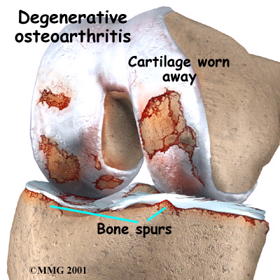
The knee joint is formed where the thigh bone, called the femur, meets the shin bone, called the tibia. That joint is covered by the kneecap or patella and is secured by muscle, tendons and ligaments, which allow the knee to bend and straighten.

The surfaces where the two bones touch are covered with a tough substance called cartilage. The space called the synovium inside the knee contains synovial fluid that helps to cushion the weight supported by the knee joint and also lubricates the bones – working like grease on a squeaky hinge.

In osteoarthritis, the synovial fluid loses its ability to lubricate ans cushion the joint effectively. As a result, the cartilage can be destroyed. The unprotected bones then rub together causing pain.
Book Appointment Online for Synvisc Treatment