Avascular necrosis (AVN) or osteonecrosis is a disease where the blood flow to the femoral head is damaged. Part of the bone of the femoral head then undergoes necrosis – it dies. When the bone dies, it becomes painful. The pain may be present either with activity or at rest. The joint surface of the femoral head may collapse and arthritis will result.
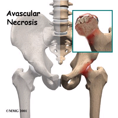
Avascular Necrosis

Symptoms usually present twelve to eighteen months after the initial damage to the bone. The cause of AVN may be traumatic such as dislocation or femoral neck fracture, or may be atraumatic and systemic.

When the cause is traumatic, a fracture disrupts the blood supply to the femoral head. Blood vessels run through the posterior capsule of the hip joint which is injured by the fracture. The blood supply to the femoral head is interrupted and AVN develops. A similar mechanism happens with traumatic dislocation. The head of the femur is forced out of the socket and the capsule is torn. Most often dislocation occurs in a posterior discretion.

Some studies show that the time from when the hip is dislocated to when it is put back in place is a factor in whether AVN develops. The shorter the time the better. AVN may develop following a femoral neck fracture even though the fracture itself heals.

Many systemic factors have been felt to lead to avascular necrosis. These include:

  • Steroid therapy
  • Alcoholism
  • Collagen vascular disease
  • Graucher’s disease
  • Arterial occlusion
  • Blood clotting disorders
  • Other diseases of circulation
  • Other blood diseases called haemoglobinopathies where small vessels in the head of the femur may be occluded.

Avascular Necrosis

Radiation injury and caisson, or dysbaric disease, are two other causes of AVN which do not fit the trauma or systemic categories. Radiation injury follows large doses of radiation given to a patient to treat other conditions. Caisson disease can occur in deep sea divers who return to the surface too quickly without allowing adequate time to decompress. With good training this rarely happens.

Patients who have had organ transplants are known to develop avascular necrosis, possibly because they medication to suppress their immune systems. In some cases, the cause of AVN may be idiopathic. This means that it develops spontaneously with no known cause.

Even though steroid therapy is one of the most common causes of AVN it is not known what dose of steroids or what length of treatment causes the disease. In systemic conditions, it is not clear if AVN occurs with a single insult to the circulation or gradually over time. It may be the result of multiple insults to the marrow in the same area.

The first symptom of avascular necrosis is pain. At the onset of the disease, the hip maintains a good range of motion. As the disease progresses, range of motion decreases. Examination and x-ray findings become similar to those in osteoarthritis. AVN is different from osteoarthritis, however, because disease first develops in the bone rather than in the joint.

On x-ray, AVN appears as a wedge of abnormal bone in the superior, or upper, part of the head of the femur. On a lateral x-ray a crescent sign may be visible. This is a clear line just below the surface of the joint. At this point the surface remains intact and has not collapsed. Within the femoral head an area of dense whitish appearing bone is visible. This indicated a process of bone repair called creeping substitution.

Often the diagnosis and surgical treatment of AVN are delayed. If pain is present and there are no changes on x-ray, the diagnosis may sometimes be made with an MRI study. Patients may delay treatment until symptoms are more severe. At the very least they should seek an orthopaedic consultation. If they wait too long, some of the more conservative options such as core decompression may no longer be helpful and hip replacement is required.

Hip replacement is not the only treatment option for avascular necrosis. If the disease is not far advanced, then other procedures may be done in an attempt to save the femoral head. This is particularly true for patients in their 30s and 40s. More conservative procedures include:

  • Core decompression with or without bone grafting
  • Bone grafting through a cortical window
  • Osteotomy
  • Resurfacing

Resurfacing may not be possible if there is too much bone collapse or there is involvement of the bone in the neck of the femur.

For older patients and for younger patients in whom the femoral head has collapsed, hip replacement is the surgical treatment of choice.
Book Appointment Online for Avascular Necrosis Treatment

Post-traumatic arthritis is arthritis of the hip joint that develops after an injury. The injury can be a severe contusion to the hip or a fracture of one of the bones within or near the joint.

A hard bruise to the hip joint or fall on the hip may not always cause a visible fracture in bone. It can, however, cause an injury to the surface or articular cartilage of the joint. The damaged surface no longer moves smoothly and the joint deteriorates and develops arthritis.

Articular injuries are hard to diagnose because the joint surfaces cannot be seen on plain x-ray. Injuries to the joint may sometimes be diagnosed on MRI. Other times, the joint injury can present as prolonged pain and stiffness with no positive radiologic findings at all. Months of years later, arthritis can develop and the severity of the joint injury is evident.

Most often post-traumatic arthritis develops after a fracture in or around the hip. Fractures through the head of the femur are unusual but can occur either as an isolated injury or in combination with a dislocation. Fractures into the hip joint socket, the acetabulum, are more common. Either of these injuries results in a damaged joint surface and can lead to post-traumatic arthritis.

Arthritis can also result from fractures that do not directly involve the joint. Examples are an intertrochanteric fracture of the hip or a fracture of the long portion or shaft of the femur. Even though the fracture heals away from the joint, the shape of the femur may change and this can lead to altered mechanics in the joint. Some parts of the joint may develop abnormal pressure and subsequent arthritis.

Since it results from injury, post-traumatic arthritis is most often unilateral or involves one hip only. Injuries to both hips are less common. They usually result from high velocity trauma such as an automobile accident.

Post-traumatic arthritis can develop following a low impact osteoporotic fracture in the elderly. The incidence is lower in these fractures because elderly people place less demand on their hips and have a shorter life span in which to develop the arthritis symptoms.

Hip replacement in post-traumatic arthritis is different than a routine primary total hip. If there has been previous surgery, there may be hardware which needs to be removed. Scar tissue may be present which makes dissection more difficult. A fracture deformity of the femur may make it harder to place standard components. A defect in the acetabulum may require a bone graft.

In general, the symptoms of post-traumatic arthritis are similar to osteoarthritis, but the surgery can sometimes be a little more difficult.
Book Appointment Online for Post-Traumatic Arthritis Treatment

Rheumatoid arthritis is a systemic disease that affects multiple joints in the body. Unlike osteoarthritis, which is due to age and wear, rheumatoid arthritis is the result of a disease process throughout the entire body. As a rule, multiple rather than single joints are affected.

Rheumatoid arthritis usually develops between ages 30 and 60 years, but can also present earlier as juvenile arthritis in childhood. Women are more than twice as likely to have the disease as men. There may be a genetic predisposition to getting the disease. Some evidence suggests that rheumatoid arthritis may be an autoimmune phenomenon, but it is unclear as to what triggers the immune response.

The source of the disease is the synovium, the lining tissue of the joint. In a healthy joint, synovium provides lubricant and nutrition to the surrounding tissues. In rheumatoid arthritis, the synovium becomes inflamed and proliferates. The resulting tissue layer is called pannus. Pannus invades the joint surfaces and causes destruction of the cartilage and underlying bone. Inflammatory factor in the synovial fluid may also damage the cartilage.

The diagnosis of rheumatoid arthritis is based largely on clinical factors, but blood tests may also be positive. The rheumatoid factor, an antibody found in blood, is present in most patients with rheumatoid arthritis. The erythrocyte sedimentation rate (ESR) is a less specific test that is often elevated in RA.

The American College of Rheumatology lists seven criteria for the diagnosis of rheumatoid arthritis. Four of these have to be positive in order to confirm the diagnosis. The seven criteria include:

  • Morning stiffness
  • Arthritis in at least three joints
  • Arthritis in the hands
  • Joint involvement on both sides of the body
  • Positive rheumatoid factor
  • Rheumatoid nodules
  • X-ray changes consistent with rheumatoid arthritis

Joints with rheumatoid arthritis are warm, swollen, and sometimes contracted. Other clinical features include rheumatoid nodules beneath the skin and tenosynovitis or thickening of the tissue that covers tendons.

The hip is one of many joints in the body that can be affected by RA. Like OA, the onset is gradual. The main symptoms are pain, stiffness, loss of motion, and limping. Usually the diagnosis of rheumatoid arthritis is known to the patient by the time symptoms present in the hip.

The surgical treatment for RA is the same as for OA. If the joint is destroyed total hip replacement is required.

Rheumatoid bone is often osteoporotic and of poor quality. This may affect the choice of components and the type of fixation. If the bone is very soft it may not be possible to press fit components and cement may be required. Patients with rheumatoid arthritis are not candidates for resurfacing because of the risk of bone collapse and failure.
Book Appointment Online for Rheumatoid Arthritis Treatment

  • If you have an mild pain or swelling at the injection site, you may want to rest and keep an ice pack on your knee for 15 to 20 minutes, or as recommended by your doctor.
  • Avoid putting a lot of strain on your knee – such as with jogging, heavy lifting, or prolonged standing – for 48 hours after the injection, or as recommended by your doctor.

How will I know When it is Time for Another Treatment with SYNVISC or Synvisc-One?

  • Since there is no known cure for osteoarthritis of the knee, and it is a chronic condition that does not go away, it is important that you continue to see your doctor regularly
  • Track your symptoms, and if you notice pain or stiffness returning, speak with your doctor about scheduling your next appointment
  • Your doctor will decide if and when it’s appropriate to repeat treatment
  • Repeat treatment with SYNVISC and Synvisc-One has been proven to be effective with a favourable safety profile.

Osteoarthritis is a chronic condition, and regular contact with your doctor can help you prevent pain from interrupting your daily activities.
Book Appointment Online for Synvisc Treatment

Brief Outline of Osteochondritis Dissecans

Osteochondritis dissecans (loose bodies in the joint) occurs when a fragment of bone adjacent to the articular surface of a joint is deprived of its blood supply, leading to avascular necrosis. This cause the cartilage to become brittle and a piece, or pieces, may break off. If the cartilage gets into the joint, it can cause pain and inflammation. The space in the ankle joint is very small and when a bone or cartilage fragment from the talus bone gets lodged in the joint, it can cause pain, swelling, and loss of movement in the ankle. These symptoms may come and go as the free-floating fragment floats in and out of the joint. Prior ankle injuries make a person susceptible to this condition, as does any blockage of blood flow to the feet.

Anatomy and Physiology for Osteochondritis Dissecans

The tarsals are the seven bones of the ankle. The two largest tarsals carry the body weight: the calcaneus, or the heel bone, and the talus, which lies between the tibia and the calcaneus. The tibia and fibula rest on top of the talus. The articular surface of the talus is covered with cartilage to cushion and protect it. There is very little blood flow to this area so repairing damage is difficult for the body. This can result in the tissue becoming brittle and breaking off. A fracture can occur on the surface of the talus, or the cartilage may become bruised from any twisting injury causing the talus to come in hard contact with the tibia and fibula.

Cause of Osteochondritis Dissecans

Loss of blood flow to the articular surface of the talus along with injury to the bone. Repetitive wear on the cartilage and bone surface of the talus. Previous ankle injury.

Signs and Symptoms of Osteochondritis Dissecans

Pain and discomfort in the joint. If the fragment becomes detached and lodged in the joint, swelling and loss of movement may occur. A catching sensation in the ankle may be felt.

Complications If Left Osteochondritis Dissecans Unattended

The loose bodies in the joint can cause scarring and additional damage if left unattended. As the joint moves and the loose body grinds against the other cartilage and bone surfaced, it will wear at these surfaces making them rough and eventually lead to arthritis.
Book Appointment Online for Osteochondritis Dissecans Treatment