Autologous Platelet Rich Plasma, a new approach for healing muscle and ligament injuries.

What is Autologous Platelet Rich Plasma Therapy?

Autologous platelet rich plasma therapy (PRP) is a promising solution to accelerate healing of tendon injuries and osteoarthritis naturally without subjecting patients to significant risk. The philosophy is to merge cutting edge technology with the body’s natural ability to heal. PRP technology was initially developed 20 years ago to aide with wound healing and blood loss. Its benefits are now applied towards the facilitation of healing tendons and cartilage. In fact, PRP has been widely used for years in Europe to treat various tendon injuries.

How does PRP procedure work?

Using the patient’s own blood, specially prepared platelet are taken and then re-injected into the affected area. These platelets release substances known as “growth factors” that lead to tissue healing. For example, when you cut yourself, the body’s natural response is to attract platelets that release growth factors and facilitate healing. By concentrating the platelets we increase the release of growth factors which promotes the healing of tendons and cartilage.

The human body has remarkable ability to heal itself and by re-injecting concentrated platelets we are facilitating the natural healing process.

Is Platelet Rich Plasma Right For Me?

For most patient PRP offers a solid, alternative treatment for those who do not wish to have surgery. An initial evaluation will determine if PRP is a viable treatment option.

What are the benefit of PRP treatment?

Patients can expect to see significant improvement in symptoms; elimination of need for more traditional treatments such as medications, cortisone inceptions or surgery; and a dramatic return for function.

How soon can I go back to regular physical activity?

This treatment is not “quick fix” and is designed to promote long-term healing. The process of PRP requires time and rehabilitation. Patients who follow a course of physical therapy are likely to see the most improvement in the affected area. PRP accelerates and shortens the recovery process in chronic injuries and new acute injuries.

Prior studies on tennis elbow have demonstrated an 81% improvement in pain scores at six months and 93% reduction in pain follow up at 1-3 years.

Through regular follow-up visits your doctor can determine when you are able to resume regular physical activities.

How do I prepare for an injection?

Patients should schedule an evaluation to determine if PRP is a viable treatment option. An MRI and/or musculoskeletal ultrasound may be done prior to the injection to insure a proper diagnosis and rule out any condition best treated surgically.

The PRP injection usually takes an hour and is performed on an outpatient basis. Do not take anti-inflammatories one week before or after the injection as this may limit the treatment benefits.

Depending on a patient’s condition, a series of 1-3 injections may be required.

How do I care of the injection site after the treatment?

Initially the procedure may cause some localised soreness and discomfort. Patients may apply ice and elevation as needed. After one week the patient will likely begin rehabilitation program with physical therapy.

What are the significant side effects?

The risks are usually uncommon.

Osteoarthritis PRP Specialist

Dr Kevin Yip Orthopaedic Surgeon Gleneagles Singapore

Dr Kevin Yip

Book Appointment Online for Platelet Rich Plasma Injection Treatment

Osteoarthritis – What is it and what symptoms would I experience?

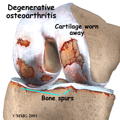
Osteoarthritis (OA) is a disease that affects the cartilage (soft tissue that protects the bone surface) in your joints, causing it to break down and wear out. Knee OA is the most common form of this disease.

Knee Osteoarthritis

Osteoarthritis:

  • Worn out cartilage
  • Decreased joint fluid

Signs and symptoms

  • Pain and tenderness at the joint
  • Stiffness (especially after getting out of bed or getting up from sitting for a long time)
  • Loss of flexibility
  • A grating feeling or the cracking sound of bone rubbing on bone.

How does knee OA develop? How do doctor diagnose it?

Knee OA develops when changes in the cartilage occur:

  • Cartilage becomes pitted, rough and brittle
  • Underlying bone thickens and broadens to reduce load on cartilage
  • Bony outgrowths form at the outer edges of the joint, making it look knobbly
  • Space inside the joint narrows. Bits of bone or cartilage can break off and float inside the joint space.

Diagnosis is usually made using several methods:

  • Medical history
  • Physical examination of the knee
  • X-rays or other imaging tests
  • Other tests such as blood tests or exams of the fluid in the joints.

What are the risks factors for OA?

  • Older age
  • Bone deformities
  • Overweight and obesity
  • Gender (women > men)
  • Joint injuries (sports or accidents)
  • Sedentary lifestyle
  • Certain occupations (that place repetitive stress on the knee)

Knee Osteoarthritis Specialist

Dr Kevin Yip Orthopaedic Surgeon Gleneagles Singapore

Dr Kevin Yip

Book Appointment Online for Knee Osteoarthritis Treatment

There is a popular belief that only middle-aged, overweight men who eat and drink too much suffer gout. This is a myth – gout can attack young men in their twenties and also women, although it is very unusual in women and occurs mainly after the menopause.

Gout Video

What is Gout Attack?

An attack of gout is caused by uric acid suddenly forming crystals inside a joint, causing intense inflammation with pain, redness and swelling. Uric acid is produced when purines, which are chemicals present within all living cells, are broken down. Purines are produced by the body itself and are also found in many foods. People prone to gout have an inherited tendency to produce a lot of uric acid. High levels accumulate in the bloodstream and are deposited in the body’s tissues.

Gouty Arthritis
Gouty Arthritis

Common sites are the joints, the kidneys and the skin over the tops of the ears, hands and elbows. If the kidneys are affected, then they do not work as well as they should at excreting the uric acid in the urine and the levels become even higher. Uric acid deposits under the skin appear as whitish lumps, called ‘tophi’. They may ulcerate and discharge material that looks like toothpaste. It is not always clear what causes the uric acid suddenly to form crystals in the joints and set off inflammation, but a common trigger is minor injury to the joint.

Gout Symptoms

A typical attack of gout is easily recognisable.

What happens during an attack of gout

  • At first, there is only minor discomfort but, within a matter of hours, the joint is swollen, hot, red and extremely painful.
  • The pain is so severe that wearing shoes is out of the question, and you may not even be able to bear the touch of a bedsheet.
  • Even with no treatment, the attack subsides completely within a few days, and always within a week.
  • Attacks may recur, although sometimes not until months or years later.
  • In 70% of people, the knuckle joint of the big toe is the first joint, and often the only joint, to be affected, although symptoms can develop in any joint.

Diagnosis

Often your doctor will be able to recognise that you have gout from your symptoms and the appearance of the affected joint. You will need to have blood tests to measure the amount of uric acid in your bloodstream and to check whether your kidneys have been affected. A sample of fluid may sometimes be taken from an acutely inflamed joint with a fine needle and syringe.

Treatment

Non-steroidal anti-inflammatory drugs are very effective and can shorten an attack if they are taken right at the beginning. Before these drugs became available, the traditional treatment for an attack of gout was a drug called colchicine, derived from the autumn crocus.

Colchicine is still used and it is also very effective, although it can cause troublesome diarrhoea in some people. In fact, both anti-inflammatory drugs and colchicine are so effective if taken early that people who have frequent attacks of gout are well advised to keep a supply in the medicine cabinet, so that they can take it at the first sign of trouble.

Diagnosing gout as a cause of joint pain

An attack of gout is caused by uric acid suddenly forming crystals within a joint, causing intense inflammation with pain, redness and swelling.

Looking ahead

If you still get frequent attacks, say more than three times a year, despite these measures, your doctor may suggest that you take drugs to lower the level of uric acid in your body. The main one is allopurinol which, taken daily as tablets, blocks the chemical pathway leading to uric acid production.

The uric acid deposits in the body tissues are then slowly removed into the urine and so leave the body.

Help yourself to avoid gout

  • Drink plenty of clear dilute fluids (water or codial), at least three litres a day, especially in hot weather and when you’re on holiday, because an attack is more likely if you become dehydrated.
  • Reduce your weight.
  • Keep your alcohol consumption within recommended levels – 21 units a week for a man and 14 for a woman. A unit is half a pint of beer, a small glass of wine or a single measure of spirits.
  • Cut down on food and drink containing high levels of purines; these include high-protein foods such as red mean and pulses, offal (liver and kidneys), sardines and anchovies. Avoid Nuts, Beans and Lentils. It is better to eat meals based around complex carbohydrates, such as pasta.
  • Avoid taking aspirin, which stops the kidneys from excreting uric acid; take paracetamol instead for minor aches and pains.

Key Points

  • Gout attacks are very painful, seek medical help early.
  • If attacks are frequent, then you may need to take tablets to reduce the level of uric acid in your body

Gouty Arthritis Specialist

Dr Kevin Yip Orthopaedic Surgeon Gleneagles Singapore

Dr Kevin Yip

Book Appointment Online for Gout Treatment

Brief Outline of Extensor and Flexor Tendinitis

The tendons attached to the muscles that are responsible for flexing and extending the toes and foot can become inflamed and irritated just like any other tendon. Overuse, tightness is opposing muscles, or foot deformities can cause this condition. Extensor tendinitis is more common that flexor, but flexor tendinitis tends to be more painful and debilitating. Dancers are most commonly associated with injury to this tendon group.

Anatomy and physiology

Foot Muscles

The extensor hallucis longus and extensor digitorum longus are the main extensor muscles of the toes. The tendons of these muscles run over the front of the ankle, over the foot and attach to the toes. These muscles dorsiflex the foot and work in opposition to the flexor muscles. When the calf muscles are tight, or the muscles are worked beyond their exertion level, inflammation of the tendon may occur.

The flexor group of muscles, the flexor hallucis longus and the flexor digitorum longus, have tendons that run down the inside of the ankle and under the foot, attaching to the toes. These muscles plantar flex the foot and toes.

Cause of Tendinitis Injury

Extensor tendinitis: Tight calf muscles, over-exertion of the extensor muscles, or fallen arches.

Flexor tendinitis: Repetitive stress to the tendon from excessive dorsiflexion of the toes.

Signs and symptoms of Tendinitis

Extensor tendinitis: Pain on the top of the foot, pain when dorsiflexing the toes, some strength loss may be experienced.

Flexor tendinitis: Pain along the tendon, in the arch of the foot, and along the inside back of the ankle.

Complications if left Tendinitis unattended

Tendinitis when left unattended can cause strains to the attached muscle and could lead to a complete rupture of the tendon. The pain may become severe enough to limit all activity.

Treatment for Tendinitis

  • Rest
  • Ice
  • Anti-inflammatory medication
  • Physiotherapy
  • Injection

Rehabilitation and prevention

While resting the foot, it is important to identify the conditions that caused the problem. Stretching the calf muscles and the tibialis anterior muscle will help relieve the pressure on the tendons. Warming-up and gradually increasing workloads will help prevent tendinitis. Orthotics may be required when returning to activity to correct any arch problems.

Long-term prognosis of Tendinitis

Most people recover completely from tendinitis with simple rest and correction of the cause(s). In some rare cases, surgery may be required to reduce the pressure on the tendons and relieve the inflammation.

Orthopaedic Specialist Treating Tendinitis

Dr Kevin Yip Orthopaedic Surgeon Gleneagles Singapore

Dr Kevin Yip

Book Appointment Online for Extensor & Flexor Tendinitis Treatment

Osteoporosis, The Silent Disease

Osteoporosis
Osteoporosis

Osteoporosis is called ‘the silent disease’ for a reason. During the early stages of the condition, you will typically have no signs and symptoms. But once your bones have been weakened by osteoporosis, you start to feel back pain, your posture will become stooped, and you will be more prone to bone fractures.

Osteoporosis is a degenerative disease that causes bone to become weak and brittle. For severe cases, even a mild fall or slight bending can cause a bone fracture. Our bone is a living tissue, which is being constantly renewed. Osteoporosis happens when the formation to new bones is slow to replace old bones, or when our body ultimately fails to form enough new bone. Although osteoporosis is sex and race blind, affecting both men and women of all races, women who are past menopause are more prone to it.

In Singapore, over the last 30 years, cases of hip fractures have increased five times in women aged 50 and above, and 1.5 times in men of the same age group. The bad news? It is a serious and debilitating condition. The good news? Osteoporosis can be prevented.

Say No To Bone Loss

Mind Your Diet

Calcium is your bones’ best friend. They help your body build strong bones. So find every opportunity to incorporate low-fat milk and non-fat yoghurt into your diet, as these are healthy sources of calcium. Fish and greens are also rich in calcium, so is soy. Sardines and salmons are your best bets, as well as brocolli, kale and bok choy.

Stop Smoking

You hear it often enought, and we say it again. Cigarette smoking is bad for your health. Cigarette smoke is packed with massive amounts of free radicals that cause a chain-reaction of damage throughout the body, including cells, organs, and hormones that work together to keep your bones strong and healthy. Amongst women, one in eight hip fractures is linked to long-term cigarette use. Studies also confirm that smokers tend to heal fractures slower than non-smokers.

Keep Active

To help strengthen bones and prevent osteoporosis, at least a 30-minute exercise daily is recommended. Exercises occupied with weight lifting help to simulate bone formation and make bone become stronger. It also helps to improve your balance and flexibility, which is necessary to reduce risks of falls that can cause fractures. Aerobics, dancing, jogging, Tai Chi, yoga, tennis, weight lifting and even climbing stairs are some of the recommendation exercises you can try.

Cut Back on Alcohol

Excessive alcohol consumption is bad for you on so many levels. When you drink too much alcohol, your body fails to absorb calcium and Vitamin D properly and adequately. As much as possible, limit your drink to one a day for women and two drinks a day for men. Cortisol and parathyroid hormones, which are both potentially bone damaging, are seen in people with alcoholism. What’s more, high alcohol intake also puts osteoblasts, the bone-making cells, at risk.

Take Bone Mineral Density Test

Osteoporosis can be prevented. Do a bone mineral density test early to check whether you are at risk of Osteoporosis.

Specialist Treating Osteoporosis

Dr Mathew Tung

Dr Mathew Tung Neurosurgeon

Book Appointment Online for Osteoporosis Treatment