The information provides advice on caring for your plaster cast, synthetic cast and splint. These applications help to immobilise an injured limb after fractures, sprains, ligament tears and other injuries.

Do’s:

  • Raise your injured part using pillows to reduce swelling.
  • Observe for change in nail bed colour of your injured part eg: from pink to blue.
  • Take painkillers when in pain.
  • Exercise your exposed fingers/toes of your injured part to prevent stiffness/swelling.
  • Keep your cast/splint clean and dry to prevent skin irritation.
  • Cover your injured part with plastic before a shower to prevent cast/splint from getting wet.
  • Use a fan to dry the inner cast/splint if it is wet or damp.

Do’s (Upper Limb)

  • Support your injured hand in an arm sling.

Arm Sling

Do’s (Lower Limb)

  • Always sit on a chair and place your injured leg on another chair when showering.
  • Use crutches/wheelchair as instructed for your own safety.

Don’ts

  • Do not put weight on the injured part unless advised by the doctor.
  • Do not wet your cast/splint.
  • Do not put powder or any object in your cast/splint.
  • Do not attempt to loosen or shorten your cast/splint.
  • Do not scratch your skin underneath your cast/splint using sharp object.

If you experience any of the following:

  • Swelling
  • Discolouration
  • Tingling sensation or numbness
  • Increasing Pain
  • Skin rubbing in the cast/splint or broken skin around the edges
  • Cast/splint feels too tight or loose, broken or cracked
  • Cast/splint is soaked or soiled

Book Appointment Online for Cast & Splint Treatment

Foot drop is the inability to lift the foot and toes properly when walking. It is caused by weakness or paralysis of the muscles that lift the foot.

People with foot drop tend to scuff their toes along the ground, so they may lift their foot higher than usual when walking to prevent this.

Foot drop is often linked to damage or disease of the brain or spinal cord, although it may also result from injury to the nerves in the leg.

If foot drop is caused by an injury or nerve damage, recovery is often possible. However, if it’s caused by a progressive neurological condition such as multiple sclerosis, foot drop will probably be a lifelong symptom to manage.

Conditions that cause foot drop

The condition causing foot drop can be temporary or permanent. Possible causes include:

  • spinal muscular atrophy
  • spinal stenosis
  • acquired peripheral neuropathy
  • damage to the nerve that passes below the knee

Managing foot drop Without Surgery

If you have foot drop, you may find it hard to clear the affected foot from the ground. This means you are more at risk of falls and injury.

Generally, making small changes in your home, such as using non-slip rugs and mats and removing clutter, can make a big difference in helping to prevent falls. Read more about preventing falls.

There are also measures you can take to stabilise the foot and improve your walking ability. These include:

  • having physiotherapy to strengthen the foot and lower leg muscles
  • wearing an ankle-foot orthosis (lower leg brace) and shoe insert
  • using an electrical stimulation device – although this is only suitable for use if the nerves controlling muscle movement are not affected

Surgery to treat foot drop

Surgery may be considered for those with permanent loss of movement from muscle paralysis. This usually involves transferring a tendon from the stronger leg muscles to the muscle that should be pulling the ankle upwards.

In rare cases, the bones of the ankle joint may be fused to stabilise the ankle.

If you are considering this form of surgery, speak to an orthopaedic specialist about what is involved and the pros and cons of the treatment.

Foot Drop Specialist

Dr Mathew Tung

Dr Mathew Tung Neurosurgeon

Book Appointment Online for Foot Drop Treatment

When diet, exercise, and medication aren’t enough,

Learn More About Targeting Osteoarthritis (OA) Knee Pain at the Source.

You Don’t Have to Settle for Osteoarthritis Knee Pain

It’s time to find the right treatment option for you

Talking to our doctor is the best way to start exploring your treatment options.

And when you do, keep in mind:

Pain affects everyone differently

The severity of your osteoarthritis (OA) as it appears on an X-ray may not reflect the amount of pain you feel. Some people with OA that appears mild on an X-ray may feel a great deal of pain. Others, whose X-rays show severe OA, may feel less pain. Your doctor is relying on you to give a clear description of how your knee feels.

Xray of the Osteoarthritis Knee

Osteoarthritis is caused by more that just cartilage loss

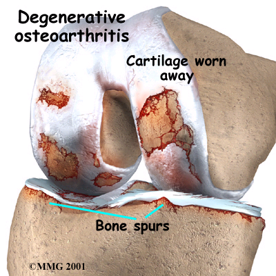
In knees with osteoarthritis, the cartilage protecting the ends of the bones gradually deteriorates, joint fluid loses its shock-absorbing qualities, and bones may begin to rub against each other – all of which may cause the knee pain you’re feeling.

Knee Osteoarthritis

It helps to know your Osteoarthritis treatment option

There are several treatment options available for osteoarthritis knee pain.

These may include:

  • Lifestyle changes. Such as exercises, physical therapy, and weight loss
  • Oral therapies, including pain relievers and nutritional supplements
  • Viscosupplement injections that provide pain relief when lifestyle changes and simple pain medications aren’t enough
  • Steroid injections in the knee
  • Surgery, such as total knee replacement

Talk to us about the risks and benefits of each treatment option to find an approach that is right for you.

Osteoarthritis Knee Specialist

Dr Kevin Yip Orthopaedic Surgeon Gleneagles Singapore

Dr Kevin Yip

Book Appointment Online for Osteoarthritis Treatment

Osteoarthritis – What is it and what symptoms would I experience?

Osteoarthritis (OA) is a disease that affects the cartilage (soft tissue that protects the bone surface) in your joints, causing it to break down and wear out. Knee OA is the most common form of this disease.

Knee Osteoarthritis

Osteoarthritis:

  • Worn out cartilage
  • Decreased joint fluid

Signs and symptoms

  • Pain and tenderness at the joint
  • Stiffness (especially after getting out of bed or getting up from sitting for a long time)
  • Loss of flexibility
  • A grating feeling or the cracking sound of bone rubbing on bone.

How does knee OA develop? How do doctor diagnose it?

Knee OA develops when changes in the cartilage occur:

  • Cartilage becomes pitted, rough and brittle
  • Underlying bone thickens and broadens to reduce load on cartilage
  • Bony outgrowths form at the outer edges of the joint, making it look knobbly
  • Space inside the joint narrows. Bits of bone or cartilage can break off and float inside the joint space.

Diagnosis is usually made using several methods:

  • Medical history
  • Physical examination of the knee
  • X-rays or other imaging tests
  • Other tests such as blood tests or exams of the fluid in the joints.

What are the risks factors for OA?

  • Older age
  • Bone deformities
  • Overweight and obesity
  • Gender (women > men)
  • Joint injuries (sports or accidents)
  • Sedentary lifestyle
  • Certain occupations (that place repetitive stress on the knee)

Knee Osteoarthritis Specialist

Dr Kevin Yip Orthopaedic Surgeon Gleneagles Singapore

Dr Kevin Yip

Book Appointment Online for Knee Osteoarthritis Treatment

Brief Outline of Claw Foot

Pes cavus (claw foot) is a genetic condition that causes a high arch, giving the foot a claw-like appearance. This often leads to tight calf muscles and pain in the forefoot. People with claw foot have difficulty finding shoes that fit properly, which can lead to other foot conditions. This condition is the opposite of flat feet but is much less common.

Claw Foot

Anatomy and physiology

Pes cavus is a condition in which there is exaggerated height of the longitudinal arch, which means that the foot is fairly inflexible due to tight calf muscles. This foot positioning puts additional stress on the metatarsal heads. The calf tendon is stretched over the heel and into the arch and the raised arch puts additional stress on the calf muscles. Those people with this genetic disorder must work to correct it or work around it in their activities.

Cause of Claw Foot

Genetic condition. Possibly secondary to contractures or disturbed balance of the muscles.

Signs and symptoms of Claw Foot

Pain in the foot, especially when walking or running. Toes may be bent.

Complications if left Claw Foot unattended

If left unattended, pes cavus can lead to chronic pain and possible injury to other structures in the foot. Foot instability is common, and could lead to strains and sprains.

Treatment of Claw Foot

  • Stretch the calf muscles and the foot.
  • Seek medical help if painful and unable to walk properly

Rehabilitation and treatment of Claw Foot

Stretching the calf muscles and the foot is the fist and most important step in rehabilitation. Finding proper fitting shoes will be important as well, which will help support the arch but also prevent injury due to the instability of the foot. Strengthening the muscles of the lower leg will also support the foot. If surgery is required, it will be important to increase strength and flexibility in the muscles that are immobilised.

Long-term prognosis of Claw Foot

When treated properly, claw foot can be corrected and the symptoms relieved. Surgery may be an option, especially when pain is severe and other treatments do not help.

Foot Specialist Treating Claw Foot (Pes Cavus)

Dr Kevin Yip Orthopaedic Surgeon Gleneagles Singapore

Dr Kevin Yip

Book Appointment Online for Claw Foot Treatment