Skin sores are characterised by the appearance of blisters, bumps or ulcers on the skin around the body.
Some common causes of skin sores include:
Underlying health conditions (e.g. eczema, diabetic leg ulcers, skin cancer)
Pressure damage (e.g. skin sores)
Infection (e.g. cold sores, chicken pox)
Burn (e.g. scald, sunburn, frostbite, chemical burn, etc)
Injury (e.g. abrasion, cut)
Other situations (e.g. insect bites or stings, food allergies, medication side effects or allergies)
Managing Skin Sores:
Skin sores are skin damage as a result of prolonged pressure on the skin coupled with friction.
Due to the pressure, blood flow to the area is reduced, causing tissue damage and sores formation.
Skin sores are more common at areas where the bones are close to the skin (e.g. ankle, back, elbow, hips or buttocks). Bed or wheel-chair bound patients are at a higher risk of developing skin sores.
Skin sores are classified into 4 stages:
Stage 1
Intact skin with discolouration, the skin is firm, soft and with slight pain.
Stage 2
A superficial open wound that resembles a ruptured fluid-filled blister.
Stage 3
A deep wound with a crater-like appearance which may expose some fatty tissues or yellowish dead tissues.
Stage 4
Extensive loss of tissues which may expose the muscles or bones, with dark dead tissues at the bottom.
TIPS:
Stage 1 and 2 skin sores could be managed at home while the higher stages require medical attention as there is an increased risk of infection.
Preventing Diabetic Ulcers
Blood sugar levels should be well controlled in a diabetic patient.
Proper fitting footwear with adequate supper is important to minimise injury.
Minor foot injuries or foot infections (e.g. fungal) should be well-managed.
To maintain a clean and healthy foot, clean the foot gently with mild soap and water, pat dry and apply moisturisers.
Diabetic patients should have their feet checked regularly by an orthopaedic surgeon.
Self-management for skin sores and diabetic ulcers
A good wound dressing (e.g. foam dressings) for skin sores or diabetic leg ulcers should minimise pain and trauma during dressing change while keeping a moist wound environment. All these factors help to create an ideal environment with minimal wound disturbance for optimal wound healing.
You should see a doctor:
If the sore does not seemed to be healing.
If there is unbearable pain or the area becomes swollen with foul odour discharge.
If you are down with fever.
If you experience other new unexplainable symptoms (e.g. nausea)
psGeneralComments Off on Surgical Nail Removal for Fungal Nail Infections
Surgical Nail Removal Overview
You may want to consider surgical nail removal for severe or recurrent fungal nail infections. The whole nail or only a portion of the nail can be removed surgically.
Surgical nail removal can be done at the clinic. The orthopaedic surgeon will give an injection in the finger or toe for local anaesthesia. He will then start by loosen the skin around the nail and then tries to separate the nail from the skin by using a tool under the nail. If a portion of the nail is infected, only the affected nail is removed.
Our orthopaedic surgeon can remove the root of the nail to prevent the nail from growing back if you have recurrent nail infection that does not seems to go away with conservative treatment.
Your finger or toe will be covered with gauze and tape once the surgery is done.
What to Expect After the Surgical Nail Removal
For the first few weeks after the surgery, keep the wound dry and clean. The wound should heal very well after a few weeks.
Is it Necessary to Have My Nail Surgically Removed?
Surgical nail removal is performed when a large portion of the nail is infected and damaged or the nail is causing you extreme pain. Majority of the cases only requires to remove a portion of the affected nail.
How Well Does Surgical Nail Removal Works?
After the infected nail has been removed, the infection can be further treated by taking oral anti-fungal medicine.
What are the Risks Involved in Surgical Nail Removal?
The risk is very small. It could be:
Pain.
Infection.
Nail grows abnormally.
What to Think About for Surgical Nail Removal?
If you have been having recurrent nail infection, you can choose to have the nail removed together with its root. Once the root of the nail is removed, there is no chance for the infected nail to grow back again.
psGeneralComments Off on Synvisc – Pure and Simple
Relieves joint pain & improves mobility to put the spring back in your step and choices back into your life!
Synvisc (sodium hyaluronate) is a treatment to provide relief from the pain of osteoarthritis (OA). Now you have an alternative to the use of pain killers such as nonsteroidal anti-inflammatory drugs (NSAIDs) or steroids.
Given by your doctor, this proven treatment reduces joint pain and improves joint mobility allowing you to resume some of the activities you once enjoyed.
Synvisc Benefits: Lubricates, cushions and protects the joint
Synvisc renews more efficient lubrication and cushioning absent from the synovial fluid of an arthritic joint. It helps to restore essential properties of the synovial fluid.
Synvisc helps to lubricate, cushion and protect the joint, bringing relief that lasts up to 6 months, from one simple injection.
Synvisc is a simple procedure
The procedure is called visco-supplementation – a simple procedure done by an orthopaedic surgeon. Your doctor injects Synvisc directly into the joint where it combines with the synovial fluid.
For many patients, relief occurs after a few days and optimum results shortly, thereafter. Unlike other treatments that may require up to five injection, Synvisc only requires a single injection.
Synvisc – Pure and Simple. Helps you get back in the game! Visit www.OrthopaedicSurgeon.com.sg
Important benefits of having Synvisc treatment
Patients can look forward to some of Synvisc advantages:
Significant pain reduction from osteoarthritis
Enhanced joint mobility and joint protection
Long term relief achieved with one single injection
Resumption of many activities you once enjoyed
Synvisc is made from a natural product
Synvisc is a highly purified, clear fluid made from a natural substance called sodium hyaluronate, which has been used in medicine for over 20 years. Synvisc has been specially formulated for single injection therapy. Your doctor can give you Synvisc by injection directly into the joint to relieve the pain of osteoarthritis at that site.
Synvisc added benefits of local treatment
Synvisc avoids the side effects often seen with traditional systemic osteoarthritis treatments
Synvisc injected into the joint at that site of discomfort restores the viscosity of the synovial fluid
Discuss Synvisc with your doctor
You and your doctor can decide if Synvisc is right for you. Ask for more information about the benefits of this pure and simple way to relieve osteoarthritis pain and enhance joint function.
How the knee joint moves
To understand how this new treatment – Synvisc works, it is important to first understand how the knee joint moves.
The knee joint is formed where the thigh bone, called the femur, meets the shin bone, called the tibia. That joint is covered by the kneecap or patella and is secured by muscle, tendons and ligaments, which allow the knee to bend and straighten.
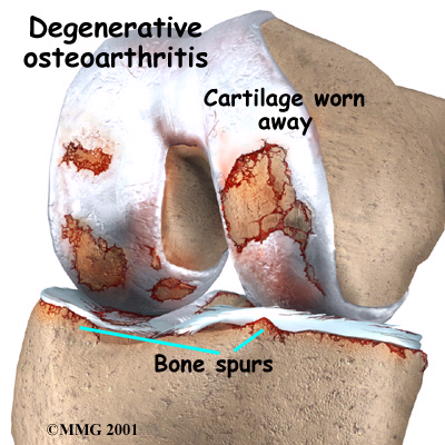
The surfaces where the two bones touch are covered with a tough substance called cartilage. The space called the synovium inside the knee contains synovial fluid that helps to cushion the weight supported by the knee joint and also lubricates the bones – working like grease on a squeaky hinge.
In osteoarthritis, the synovial fluid loses its ability to lubricate ans cushion the joint effectively. As a result, the cartilage can be destroyed. The unprotected bones then rub together causing pain.
Singapore Orthopaedic Surgeon introduces a state-of-the-art MRI scanner that has a wider opening, light-weight coils and other exciting advanced technology that improves your experience during the examination.
In addition to enhancing the experience, the Optima MR450w also provides our surgeon with excellent image quality and advanced tools to aid in their evaluation.
About Magnetic Resonance Imaging
An MRI scanner uses a powerful magnet and radio waves linked to a computer to create remarkably clear and detailed images of the body. MR imaging uses no ionising radiation, which is the radiation used in x-ray and CT machines.
MRI can be used to view, monitor, or diagnose disease abnormalities in many different regions of the body.
Benefits of Using A Wide-bore MRI
A wider opening and a shorter magnet makes the procedure more comfortable for you, especially children, those with claustrophobia and larger individuals
An exclusive optical RF system increases signal clarity and maxmises signal intensity for clean, crisp images, which leads to more accurate diagnosis and less ambiguity
Advanced functionality gives the radiologists the tools they need to make definitive diagnosis
Exclusive ease-of-use features help make life easier for both patients and the technologists
With a 50cm field of view, the technologists can cover more anatomy in fewer scans, leading to a shorter examination time
Musculoskeletal MR Imaging
MRI of the musculoskeletal system is a very valuable for evaluating joints of the body including hips, wrists, and hands. It is useful for diagnosing back and neck pain and evaluating soft tissue found near and around bones. MRI is extensively used to diagnose sports-related injuries, as well as work-related disorders caused by repeated strain or trauma.
Preparation Before Your Examination
Do inform the technologist (the person performing the MRI) if you have any of the following condition prior to the scan:
Also known as as stenosing tenosynovitis, trigger fingers are painful conditions of the fingers and thumbs where they get stuck in a bent position and then straighten with a “click” or a snap – kind of like a trigger being pulled and released. If a trigger finger is severe, you may find your finger locked in a bent position, unable to straighten.
What happens is the protective covering, the tendon sheath that surrounds the tendon of the affected finger, becomes narrow because of inflammation. This usually happens to people who have work or hobbies that require repetitive gripping actions, though they are more common in women and anyone who has diabetes.
How would you know you have trigger fingers?
Most of the time, trigger fingers or thumbs happens more frequently in your dominant hand, and mostly affects the thumb, index or ring finger. Sometimes, more than one finger is affected at the same time, and sometimes fingers on both hands may be involved. Triggering incidents are usually more often in the morning, or firmly gripping an object or when you bend and straighten the affected finger.
The common tell-tale signs and symptoms of trigger finger, in progression from mild to severe:
Stiffness in the finger, especially in morning or after periods of rest
A clicking and snapping sensation as you bend and straighten your finger
A “bump” (nodule) that is sensitive or tender at the base of the affected finger
Finger “catches” or “locks” frequently, followed by suddenly popping straight again
Finger becomes locked in a bent position, and you’re unable to straighten without pain
When you should see a doctor?
If you find that you have any of the signs above, for example, stiffness, tenderness at the base of your finger or catching in your finger joints, visit your doctor as soon as you can so that he or she can review your symptoms and do a physical evaluation of your hand. If you find that your finger joint is hot and swollen, get medical care as soon as you can, as those signs indicate possible infection.
What are the main causes of trigger fingers?
The culprit is the narrowing of the tendon sheath that surrounds the tendon in the affected finger. Tendons are thick, fibrous cords that attach muscles to bones; and each tendon is surrounded by a tendon sheath, which is lined with a substance called tenosynovium. This functions as a disperser of the lubrication which allows the tendon to glide smoothly in the protective sheath as you bend and straighten your finger, like a rope through a lubricated container.
But if the tenosynovium becomes inflamed often or for long periods, the space inside the tendon sheath becomes tight, making it difficult for the tendon to glide. The “catch” is when the tendon kinks in a bent position before popping straight . Every time the tendon kinks and snaps, the tendon becomes more irritated and inflamed, further worsening the problem. If the problem is prolonged and more inflammation occurs, the tendon can develop scars and thicken (fibrosis), and cause the tender bumps (nodules) to form.
Testing and diagnosing trigger fingers
It often doesn’t require in-depth or elaborate testing to confirm diagnosis of trigger finger. Usually your doctor will make the diagnosis based on your medical history and a hand physical exam. During the physical exam, your doctor will ask you to bend or straighten your finger, check the base of your finger for tender bumps / nodules, how smooth your finger move, and look out for the evidence of joint catching.
Treatment for trigger fingers
Avoiding repetitive gripping. For at least four to six weeks, avoid activities that require repetitive gripping or any forms of movements or activities that aggravate the problem, repeated grasping or the prolonged use of vibrating hand-held machinery.
Splinting. Your doctor may require you to wear a finger extension splint for up to six weeks to protect the injury and allow the tendon and the tendon sheath to heal. It’s especially useful to prevent you from bending your finger when you sleep, which is often painful enough to wake you up.
Finger exercises. Protected exercises – Be seated comfortably, with your hand supported. Use your unaffected fingers to bend the affected finger joints without causing the joint to click and snap. Repeat for five minutes. Gently massage the nodule at the base of your finger for five minutes. You may be referred to a hand therapist who will teach you protected mobilisation exercises to maintain mobility in your finger.
Anti-inflammatory injection. Another faster and effective way is to inject anti-inflammatory medication to the affected tendon sheath. The medicine will help to reduce the inflammation and swelling of the tendon sheath gradually.
Surgery. If your trigger finger is severe and does not respond well to conservative treatment. Surgery – trigger finger release may be another option. It is a minor day surgery and it is done under local anaesthesia.
See a doctor to treat your trigger finger
If you find that preventive or self-management measure aren’t working, see a doctor as soon as possible. Prompt medical attention can mean the difference between a quick recovery and a lifetime of suffering. And, by all means, be sure the doctor you choose is familiar with these disorders.
Who are at risk for trigger fingers?
There are three main risk factors:
Repeated gripping – if your work or hobbies require you to routinely grip objects, such as power tools or musical instruments for a prolonged duration, you may be more prone to trigger fingers.
Certain health problems – you have higher risks if you have certain medical problems such as rheumatoid arthritis, diabetes, amyloidosis, and certain infections.
Gender – trigger fingers are more prevalent in women than in men.