What are warts, and what causes them?

A wart is a skin growth caused by some types of the virus called the human papillomavirus (HPV). HPV infects the top layer of skin, usually entering the body in an area of broken skin. The virus causes the top layer of skin to grow rapidly, forming a wart. Various types of this virus thrive in warm, damp environments such as showers, locker room floors, and swimming pool areas.

Warts can grow anywhere on the body, and there are different kinds. For example,common warts grow most often on the hands, but they can grow anywhere. Plantar warts grow on the soles of the feet.

You are most likely to develop a wart where you have broken skin, such as a cut, a hangnail, a closely bitten nail, or a scrape. Plantar warts are common in swimmers whose feet are not only damp and softened but are also scratched and broken by rough pool surfaces.

Plantar Warts
Plantar Warts

How are warts spread?

Warts are easily spread by direct contact with a human papillomavirus. You can infect yourself again by touching the wart and then touching another part of your body. You can infect another person by sharing towels, razors, or other personal items. After you’ve had contact with HPV, it can take many months of slow growth beneath the skin before you notice a wart.

It is unlikely that you will get a wart every time you come in contact with HPV. Some people are more likely to get warts than others.

What are the symptoms for warts?

Warts come in a wide range of shapes and sizes. A wart may be a bump with a rough surface, or it may be flat and smooth. Tiny blood vessels grow into the core of the wart to supply it with blood. In both common and plantar warts, these blood vessels may look like dark dots in the wart’s center.

Warts are usually painless. But a wart that grows in a spot where you put pressure, such as on a finger or on the bottom of the foot, can be painful.

How are warts diagnosed?

A doctor usually can tell if a skin growth is a wart just by looking at it. Your doctor may take a sample of the wart and look at it under a microscope (a skin biopsy). This may be done if it isn’t clear that the growth is a wart. It may also be done if a skin growth is darker than the skin surrounding it, is an irregular patch on the skin, bleeds, or is large and fast-growing.

How are warts treated?

Most warts don’t need treatment. But if you have warts that are painful or spreading, or if you are bothered by the way they look, your treatment choices include:

  • Using a home treatment such as salicylic acid or duct tape.
  • Removing the wart with surgery.

Effective wart treatment will be removing it surgically. The goal of wart treatment is to destroy or remove the wart totally.

When To Call a Doctor

See your doctor if the wart is:

  • Painful.
  • Easily irritated.
  • Growing or spreading to other parts of your body or to other people.

Book Appointment Online for Plantar Wart Treatment

Brief Outline of Achilles Tendon Strain

Achilles tendon strains can be very painful and take some time to heal. The Achilles tendon, which gets its name from the mythological Greek warrior Achilles, is located in the back of the lower leg over the heel. An injury to this tendon can be debilitating because of its involvement in walking and even balance during weight bearing. Explosive activities such as sprinting and jumping, and those activities that involve pushing against resistance such as football linemen and weight training, contribute greatly to this injury.

Achilles Tendon Rupture

Anatomy and Physiology for Achilles Tendon

The Achilles tendon is the largest tendon in the human body, being approximately 15 cm long, and 2 cm thick. It originates from the musculo-tendinous junction of the calf muscles, and inserts into the posterior aspect of the calcaneus. The tendon is separated from the calcaneus by the retrocalcaneal bursa, and from the skin by the subcutaneous calcaneal bursa. It pulls the foot downward, extending it when the calf muscles contract. The strain can be graded on a scale from 1 to 3.

Grade 1 strain: A stretching or minor tear of the tendon (less than 25% of the tendon.)

Grade 2 strain: Involves more of the tendon fibres (usually 25% to 75%.)

Grade 3 strain: A complete rupture of the tendon.

Ruptured Achilles Tendon


Cause of Achilles Tendon Strain

Abrupt, forceful contraction of the calf muscles; especially when the the muscle and tendon are either cold or inflexible. Excessive force applied to the foot, forcing the foot into the plantar flexion.

Signs and Symptoms of Achilles Tendon Strain

Pain in the Achilles tendon, from mild discomfort in grade 1 strains to severe, debilitating pain in the grade 3 strains. Swelling  and tenderness may also be experienced. Pain when rising on the toes. Stiffness in the calf and heel area after resting.

Complications If Left Achilles Tendon Strain Unattended

A minor tear may become a complete rupture if left unattended. Bursitis and tendinitis may develop from the inflamed tendon rubbing over the heel.

Immediate Treatment to Achilles Tendon Strain

R.I.C.E. Anti-inflammatory medication. Then Shockwave Therapy to promote blood flow and healing. Immobilisation and medical help for complete ruptures.

Rehabilitation and Prevention for Achilles Tendon Strain

Rest is important and a gradual return to activity must be undertaken. Stretching and strengthening the calf muscles is important in rehabilitation and to prevent a recurrence. Warming-up the calf muscles properly before all activities, especially those involving forceful contractions such as sprinting, is essential to prevent strains.

Long-term Prognosis for Achilles Tendon Strain

Due to lower blood supply in tendons, they take longer to heal than the muscle, but with rest and rehabilitation, the Achilles tendon can return to normal function. Complete ruptures occasionally require surgical repair.
Book Appointment Online for Achilles Tendon Strain Treatment

Achilles Tendinitis Video

Brief Outline of Achilles Tendinitis

Inflammation of the Achilles tendon can be very painful, especially since all of the body’s weight is supported by this structure and the shoe often presses against this area. Repetitive stress to the tendon can lead to inflammation that causes additional irritation, causing more inflammation. Activities such as basketball, running, volleyball, and other running and jumping sports can lead to Achilles tendinitis.

Anatomy and Physiology of Achilles Tendinitis

The Achilles tendon is the largest tendon in the human body, being approximately 15 cm long, and 2 cm thick. It originated from the musculo-tendinous junction of the calf muscles and inserts into the posterior aspect of the calcaneus. The tendon is separated from the calcaneus by the retrocalcaneal bursa, and from the skin by the subcutaneous calcaneal bursa. The tendon crosses the back of the heel, which means it rides over the bone as the muscle contracts and stretches. Repetitive contraction of the muscles in the calf and improper footwear or excessive pronation of the feet can lead to inflammation in the tendon.

Achilles Tendinitis

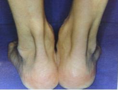
Right Foot Tendon is Thicker than Left Foot Tendon

Cause of Achilles Tendinitis

Repetitive stress from running and jumping activities. Improper footwear or awkward landing pattern of the foot during running. Untreated injuries to the calf or Achilles tendon.

Signs and Symptoms of Achilles Tendinitis

Pain and tenderness in the tendon. Swelling may be present. Contraction of the calf muscle causes pain; running and jumping may be difficult.

Complications If Left Achilles Tendinitis Unattended

Inflammation in the tendon can lead to deterioration of the tendon and eventual rupture if left untreated. Inflammation may lead to tightening of the tendon and attached muscle, which could lead to tearing.

Immediate Treatment for Achilles Tendinitis

Rest, reducing or discontinuing the offending activity. Ice. Anti-inflammatory medication. Shockwave therapy to promote blood flow and healing.

Rehabilitation and Prevention of Achilles Tendinitis

After a period of rest, usually lasting 5-10 days, gentle stretching and strengthening exercises can be initiated. Heat may be used on the tendon before activity to warm the tendon properly. Adequate warm-up, along with strengthening and stretching exercises for the calves, will help prevent tendinitis of the Achilles tendon.

Long-term Prognosis for Achilles Tendinitis

Tendinitis seldom has lingering effects if treated properly. Tendinitis may take from five days to several weeks to heal, but rarely needs surgery to repair it.
Book Appointment Online for Achilles Tendinitis Treatment

Brief Outline of Ankle Sprain

Anyone involved in athletics is susceptible to an ankle sprain, an acute injury to any or all of the ligaments that support that support the ankle structure. Tearing or stretching of the ligaments can occur when the foot is rolled either medially or laterally, or twisted forcefully. High impact sports involving jumping, sprinting or running on changing or uneven surfaces often lead to ankle sprains. Basketball, football, cross country, and hockey are a few of the sports commonly associated with ankle sprains.

Anatomy and Physiology of Ankle Sprain

Lateral ankle sprains commonly occur when stress is applied to the ankle during plantar flexion and inversion, injuring the anterior talofibular ligament. The medial malleolus may  act as a fulcrum to further invert the calcaneofibular ligament if the strain continues. The peroneal tendons may absorb some of this strain. Medial ankle sprains are less common, because of the strong deltoid ligament and bony structure of the ankle. The ligaments are stretched beyond their normal range and some tearing of the fibres may occur. Forceful twisting or rolling of the ankle, as with landing on the outside of the foot, can stretch the ligaments past their stretch point.

Cause of Ankle Sprain

Sudden twisting of the foot. Rolling or force to the foot, most common laterally.
Ankle Sprain

Signs and Symptoms of Ankle Sprain

First-degree sprains: Result in little or no swelling, mild pain, and stiffness in joint.

Second-degree sprains: Commonly exhibit more swelling and stiffness, moderate to severe pain, difficult with weight bearing,  and some instability in the joint.

Third-degree sprains: Result in severe swelling and pain, inability to bear weight, instability in the joint, and loss of function in the joint.

Complication if Left Ankle Sprain Unattended

Chronic pain and instability in the ankle joint may result if left unattended. Loss of strength and flexibility, and possible loss of function may also result. Re-injuring the joint is much more likely as well.

Immediate Treatment for Ankle Sprain

R.I.C.E. Second- and third-degree sprains may require immobilisation and should seek medical attention immediately.

Rehabilitation and Prevention for Ankle Sprain

Strengthening the muscles of the lower leg is important to prevent future sprains. Balance activities will help to increase proprioception (the body’s awareness of movement and position of the body), and strengthen the weakened ligaments. Flexibility exercises to reduce stiffness and improve mobility are needed also. Bracing during the initial return to activity may be needed but should not replace the strengthening and flexibility development.

Long-term Prognosis for Ankle Sprain

With proper rehabilitation and strengthening, the athlete should not experience any limitations. A slight increase in the probability of injuring that ankle may occur. Athletes who continue to experience difficulty with the ankle may need additional medical interventions, including, in rare cases, possible surgery to tighten the ligaments.
Book Appointment Online for Ankle Sprain Treatment

Brief Outline of Ankle Fracture

The ankle joint is one of the most commonly injured joints in the body. The majority of athletes have experienced at least a minor sprain of the ankle. Ankle fractures are less common, but nonetheless more common than other fractures. Due to the ankles involvement in all running and jumping activities, it is very susceptible to injury. Running or jumping on uneven or changing surfaces can lead to ankle fractures. High impact sports such as football and rugby, where the possibility of forceful twisting of the ankle may occur, also have a high incidence of ankle fractures.

Anatomy and Physiology of Ankle Fracture

Ankle Fracture

The ankle joint, is a hinge joint, and comprises the tibia, fibula, and talus bones. The ankle joint articulates between the distal tibia, the medial malleolus of the tibia, the lateral malleolus of the fibula and the talus. These bones are held together by a series of ligaments. In an ankle fracture, any or all of the bones and ligaments may become involved. Ankle fractures most commonly involve the ends of the tibia or fibula, or both, with some ligament stretching and tearing present as well.

Cause of Ankle Fracture

Forceful twisting or rolling of ankle can cause the end of the bones to fracture. Forceful impact to the medial or lateral side of the ankle while the foot is planted.

Signs and Symptoms of Ankle Fracture

Pain to touch. Swelling and discolouration. Inability to bear weight. Deformity may be present in the ankle joint.

Complication If Left Ankle Fracture Unattended

An ankle fracture that is left unattended can result in incorrect or incomplete healing of the bones. Continued walking or running on the injured ankle could result in further damage to the ligaments, blood vessels, and nerves that pass through the joint.

Immediate Treatment for Ankle Fracture

Stop the activity. Immobilise the joint and apply ice. Seek medical attention.

Rehabilitation and Prevention of Ankle Fracture

While the ankle is immobilised, it is important to keep conditioning levels up by using upper body exercises and weight training. When cleared for the activity with the ankle, strengthening and stretching of the muscles of the lower leg is essential for a speedy recovery. An ankle brace may be needed for support during the initial return to activity. Stronger calf and anterior muscles help support the ankle and prevent or lessen the incidence of injuries. Avoid running and jumping on uneven surfaces as much as possible.

Long-term Prognosis for Ankle Fracture

Although people who have fractured their ankle tend to have a slightly higher rate of re-injury, proper strengthening and rehabilitation usually lead to a full recovery. Compound fractures or those in misalignment may require surgical pinning to hold the bone in place while it heals.
Book Appointment Online for Ankle Fracture Treatment