Lower Back Pain

Diagnosing and Treating Back Muscle Injuries

If in addition to pain, you have weakness, bowel or bladder problems, numbness, or tingling in your arms or legs, these suggest that you have more than a muscle problem and indeed suggest a disc herniation, broken vertebra, or spinal instability, resulting in the compression of the cord or nerve roots. For moderate-to-severe pain, see your doctor immediately. For what you interpret as just muscle pain, see your doctor if it persists for a month or so.

We can’t see a spasm on X-ray or MRI, and it may not be detectable by many sophisticated diagnostic tests, but your doctor can diagnose it by touching the muscle in question and feeling the tightness.

High-quality tests like an MRI is referred to see if there is damage to a spinal disc, a fracture or slipped disc.

You can Ease the pain with:

  • Medication
  • Physiotherapy
  • Injection

What is Muscle Spasm?

Muscle spasm is a common cause of acute back and neck pain. Many backs go into spasm following overuse, playing sports without warming up first may strain their backs. Others lift or pull heavy objects using their backs rather than their legs for leverage and cause a spasm. There are many ways to hurt the muscle around the spine as there are people who put themselves out of action with this type of acute back pain. Muscles in any part of the spine can go into spasm, as it is for disc herniations.

Herniated Disc

A muscle spasm is an involuntary local muscle contraction. It can be acute or chronic. It can be caused by repetitive overuse: bending the back for long hours. Or it can be caused by misuse and muscle injury during sports. It can also occur in response to an underlying painful problem, such as an acute disc herniation or chronic pain following failed spinal surgery.

Any awkward movement – misuse or injury – can lead to a severe , painful muscle spasm and even cause the back to lock in place. Not only exertion; ordinary things like sneezing, coughing, bending to tie your shoe, or turning to face a different direction can cause a spasm. Finally, many of you who have had disc herniation know how some of the muscles in the back or neck, occasionally in the leg or shoulder, go into painful spasm.

Acutely damaged muscles become painful because acid and other toxic chemicals are produced within the muscle after overuse: a reaction to misuse or injury. Any spasm, regardless of cause, can turn the muscles of the back into a hard, painful knot. You have likely has a severe back or neck spasm or know of someone who has; it literally keeps you from moving, as if you were in painful, muscular cast.

Not only do injured muscles often go into spasm but chronically painful underlying spinal conditions, such as disc herniation, can cause chronic spasm, worsening the underlying pain. Here the spasm is mediated by the nervous system; it is not due to muscle injury. Such a reflexive spasm is caused by excessive pain impulses travelling up the root compressed by the disc to the cord. Then, through a reflex mechanism there, other nerve impulses are sent back to the root, which results in spasm of the muscles supplied by it.

This reflex mechanism may be active even if the root is not compressed or irritated. For example, the pain from a broken vetebra or one damaged by a tumour may cause spasm of the muscles around it. In this case, both the vertebra and the affected muscles are supplied by the same roots. In these cases of reflex spasm, our muscles have formed a sort of splint to protect the underlying spine. Unfortunately, the splint may be as painful the underlying problem.

Ongoing spasm, regardless of the cause, injures the muscle to some extent. However, once the underlying cause is removed, the spasm should diminish in at most a few days.

Straining Your Back

The difference between a strain and spasm may be academic when it comes to sore back muscles, since the treatment is similar. Strain is caused by predominantly by overuse: repetitive, forceful movements that result in muscle soreness without spasm. Muscle is made up of individual and segmental strands of tissue. When any muscle, including those around the spine, encounter excessive pulling force, individual strands can stretch or tear – become strained – while the rest of the muscle remains intact. If you do lots of bending, lifting, and twisting, you are probably no stranger to this type of back pain. On the opposite side of the coin are people who rarely exercise. Muscles and ligaments become weak. When an inactive person suddenly becomes active, injuries are common.

The major symptom of back strain is pain when you stretch the muscle and you feel it tighten. If the strain is more severe, the symptoms are abrupt and immediately disabling. They are often accompanied by what patients describe as an audible snap or pop. After the initial  burst of pain, symptoms of severe strains may subside, making them sometimes less painful than minor strains. Lumbar muscle strains may cause a broad, aching pain across the lower back or pain may be limited to one side. You may have trouble bending down or stranding up straight. You may also feel an occasional spasm when you move around or even while sleeping.

Prevention

After you have sprained a back muscle, it is more vulnerable to another injury.

  • Exercise regularly to strengthen your back and abdominal muscles, which support your spine.
  • Good posture.
  • Use ergonomics.

Muscle Spasm and Back Pain Specialist

Dr Mathew Tung

Dr Mathew Tung Neurosurgeon

Book Appointment Online for Muscle Spasm & Back Pain Treatment

Back pain is an extremely common complaint. 90% of the adult population would have experienced back pain at some point in their lives and approximately half of these people would have experienced recurrent back pain.

Many conditions give rise to back pain. The most common source of pain is related to the encountered causes include disorders of abdominal oragans (pancreatitis, kidney stones or infection), blood vessels injuries (aortic dissection) and infections affecting nerves (shingles). Despite investigations, some cases of back pain have not attributable cause. The good news is that, no matter what the cause of the pain is, the majority of back pain resolves spontaneously within six to eight weeks.

Spine Anatomy Top ViewCauses of Back Pain

The following are common causes of back pain related to the spine and surrounding structures.

Degenerated lumbar disc

A lumbar disc is the soft tissue found between each vertebra. It is responsible for spinal stability, mobility and absorption of shock transmitted through the spine. 80% of the composition of a normal disc is water. This gives it a springy consistency and allows for normal function. When a disc undergoes degeneration, it loses some of its water content. As a consequence of this, the disc loses its thickness and bulges at its periphery. This bulging irritates nerves found at the edge of the disc, giving rise to back pain. This pain can radiate down to the buttocks and is aggravated by prolonged sitting, one would tend to slouch and this flexes the spine, increasing the pressure on the disc. This further irritates the nerve, thus worsening the pain in the back and buttocks (discogenic back pain).

Herniated lumbar disc (slipped disc)

Herniated Disc

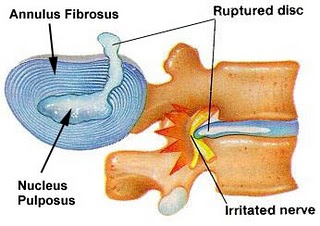
A herniated lumbar disc occurs when the central jelly-like part of the disc (nucleus pulposus) bulges through a fissure in the outer capsule (annulus fibrosis) of the disc. This herniation often causes pressure on the nerves supplying the lower limbs resulting in leg pain, in addition to back pain. There can also be lower limb weakness and numbness due to impairment of nerve function secondary to compression. Occasionally, the herniated disc can cause compression of the nerves supplying the bowel and bladder resulting in incontinence (cauda equina syndrome). This is an emergency condition which requires urgent surgery. If there is a delay in the treatmetn of cauda equina syndrome, incontinence can be permanent.

Spinal Stenosis

Spinal stenosis is characterised by back pain associated with leg pain after walking for a certain distance. This phenomenon is known as neurogenic claudication. The walking distance achievable is predictable and the leg pain can also be predictably relieved by sitting down but not by standing still. This condition is caused by narrowing of opening of the spine through which the nerves supplying the lower limbs exit. The narrowing is often due to arthritis of the small joints of the spine (facet joints) which results in enlargement of the joints from growth of bone spurs (osteophytes). The narrowing is also contributed by bulging, degenerate discs.

Myofascial pain

This is pain originating from the muscles and supporting soft tissues surrounding the skeletal portion of the spine. It is characterised by tenderness over localised areas  on the back (trigger points). Pain is typically relieved by stretching the involved muscle group or by trigger point release (massage) or injection.

Spinal Fractures, Infections and Tumours

While uncommon, these are particularly sinister causes of back pain associated with poor outcomes if not detected and treated early.

When to Seek Treatment

These are factors that may point towards a condition that requires urgent treatment such as a fracture, infection or tumour (“red flags”).

  • Recent trauma such as a fall from a height or motorcycle accident.
  • Recent mild trauma in an individual above the age 50 such as a fall onto the buttocks.
  • History of prolonged steroid use.
  • Someone with a history of osteoporosis.
  • Any person above the age of 70 due to increased risk of cancer, infection and non-spinal cause of back pain.
  • Previous history of cancer.
  • History of recent infection.
  • Temperature over 38.5 degrees Celsius associated with back pain.
  • Intravenous drug use.
  • Back pain worse at rest which is associated with infections and tumours.
  • Recent unexplained weight loss.

Other characteristics of pain which warrant urgent medical attention include –

  • Prolonged back pain.
  • Back pain associated with acute onset weakness or numbness of the lower limbs or lower limb pain.
  • Back pain associated with incontinence.

Assessment and Treatment

A doctor’s assessment of back pain includes obtaining a history regarding the pain, associated symptoms and conditions. This is followed by an examination of the back and nerve function of the lower limbs.

If a spinal condition for back pain is suspected, a X-ray will be performed. A MRI scan may also be requested for if there is any impairment of nerve function or presence of any “red flags”.

The majority of back pain is treated with rest, medications and physical therapy. In very selected conditions, surgery is necessary.

Back Pain Specialist

Dr Mathew Tung

Dr Mathew Tung Neurosurgeon

Book Appointment Online for Back Pain Treatment

Scoliosis is a problem with the curve in your spine. Some curves in your spine are normal. But a few people have spines that make a large curve from side to side in the shape of the letter “S” or the letter “C.” If this curve is severe, it can cause pain and make breathing difficult.

The good news is that most cases of scoliosis are mild. If found early, they can usually be prevented from getting worse.

Scoliosis Bending Down

Scoliosis

What causes scoliosis?

Scoliosis usually starts in childhood. Scoliosis that is severe enough to need treatment is most common in girls.

A curve in the spine may get worse as your child grows, so it is important to find any problem early.

In most cases, the cause of scoliosis is not known. This is called idiopathic scoliosis. It develops mostly in children and teens and appears to be related to several things, including genetics, as it often runs in families.

There are two types of scoliosis: nonstructural and structural.

Nonstructural (functional) scoliosis

Nonstructural (functional) scoliosis involves a curve in the spine, without rotation, that is reversible because it is caused by a condition such as:

  • Pain or a muscle spasm.
  • A difference in leg length.

Structural scoliosis

Structural scoliosis involves a curve in the spine, with rotation, that is irreversible and is usually caused by an unknown factor (idiopathic) or a disease or condition such as:

  • Disorders that were present at birth (congenital), such as spina bifida, in which the spinal canal does not close properly; or a disorder that affects the formation of bones. These curves can be harder to correct. They often get worse as the child grows, especially during the teen years.
  • Nerve or muscle disorders, such as cerebral palsy, Marfan’s syndrome, or muscular dystrophy.
  • Injuries.
  • Infections.
  • Tumors.

In adults, scoliosis may result from changes in the spine due to aging (degenerative changes). These degenerative changes may be caused by osteoarthritis.

What are the symptoms?

Scoliosis most often causes no symptoms in your child until the spinal curve becomes large. You might notice these early signs:

  • Your child has one shoulder or hip that looks higher than the other.
  • Your child’s head does not look centered over the body.
  • Your child has one shoulder blade that sticks out more than the other.
  • Your child’s waistline is flat on one side, or the ribs look higher on one side when your child bends forward at the waist.

In adults, scoliosis may cause back pain and trouble breathing.

How is scoliosis diagnosed?

The doctor will check to see if your child’s back or ribs are even. If the doctor finds that one side is higher than the other, your child may need an X-ray so the spinal curve can be measured.

Scoliosis is most serious in young children who are still growing. A curve in the spine may get worse as your child grows. So many experts believe screening your child for scoliosis is important so that any curve in the spine can be found early and watched closely.

How is Scoliosis treated?

Mild cases of scoliosis usually do not need treatment. Your doctor will check the curve of your child’s spine every 4 to 6 months. If the curve gets worse, your child may need to wear a brace until he or she has finished growing. In severe cases, or if bracing doesn’t help, your child may need to have surgery.

Scoliosis and its treatment can be a severe strain on your child. Wearing a brace can feel and look odd. It also limits your child’s activity. Your child needs your support and understanding to get through treatments successfully.

What increases the risk of scoliosis?

Your child may be more likely to have scoliosis if someone in your family has had it and if your child is a girl. Other things that increase the chance of scoliosis include:

  • One of the bones in your child’s spine has moved forward out of place compared to the rest of the spine.
  • Your child’s arms or legs are missing or are abnormally short.
  • Your child has other problems with tissue growth that happened before birth.

When To Call a Doctor

Call your doctor to have your child evaluated for scoliosis if:

  • You observe a curve in your child’s spine.
  • You notice that something about your child’s posture looks unusual, such as ribs that stick out, one shoulder that is higher than the other, one hip that is higher than the other, or an uneven waistline.
  • You observe that your child’s clothes don’t fit properly or that his or her hems don’t hang evenly.
  • A school screening program recommends that your child see a doctor.

Book Appointment Online for Scoliosis Treatment

The lumbar disc is comprised of an inner gel and an outer annulus. The lumbar discs provide stability and resiliency to the lumbar spine. Lumbar disc herniation develops because of a tear in the annulus of the lumbar spine; the inner nucleus pulposus extends beyond the tear, comprising a part of the spinal canal.

Prolapsed Intervertebral Disc

In many cases, lumbar disc herniation develops insidiously as a result of repeated, small micro tears that develop in conjunction with a degenerative disc. In these cases, patients with lumbar disc herniation may be completely without symptoms. Indeed, imaging studies have revealed that lumbar disc herniation may be present in a sizable number of individuals who have no symptoms.

Many patients with an acute lumbar disc herniations have symptoms of pain or numbness in conjunction with a pinched or compressed nerve. Acute lumbar disc herniation develops because of a sudden and large tear in the annulus, with subsequent rupture of nucleus pulposus material through the tear. Most of these herniations occur posteriorly, that is, toward the back. They also are generally to one side, but occasionally are midline.

If a disc herniation is initially midline, pain is perceived as a deep discomfort in the center of the lower back. Sometimes patients describe a sudden popping sensation. Other times, the patient senses that something is wrong in the lower back, but the pain may not be excruciating. Over days, the tear may enlarge and the disc herniation worsens while symptoms increase.

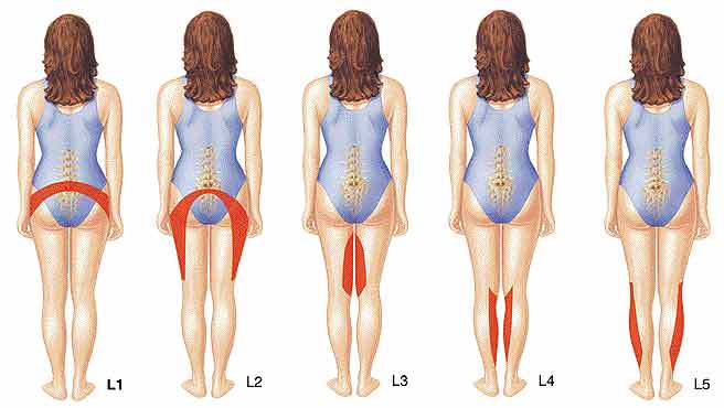
As a disc herniation enlarges, it generally does so to the right or left of midline. Often, it will compress the lumbar nerve root at that level. Most disc hernations develop between L4-L5, or L5-S1, which are the two lowest segments of the lumbar spine.

These two levels of the lumbar spine are the most mobile and vulnerable segments.

lumbar_herniated_prob_areas

When a nerve root is compressed, there may be associated pain, and pain will normally be perceived in the sensory distribution of that nerve. For example, a herniated disc at L4-5 may compress either the right or left L5 nerve root, which may cause pain radiating down the right or left side of the leg and into the great toe. IF the nerve is severely compressed, there may be numbness (corresponding to the same sensory distribution) or muscle weakness. This demands urgent medical attention.

Patients with acute lumbar disc herniation typically are comfortable lying on their back with knees flexed. Sitting is often uncomfortable, and standing may or may not be comfortable. Patients generally avoid bending forward, and sudden coughing or sneezing can worsen symptoms.

Cause of Lumbar Disc Herniation

The cause of acute lumbar disc herniation is not always apparent. Patients with chronic, degenerative lumbar discs and associated disc herniations often have a genetic predisposition to such degenerative changes. Otherwise, lumbar disc herniations generally occur in the setting of repeated overuse of the lumbar spine or sudden, more severe overuse. In this case, it is not simply the supporting musculature that gives, but the stabilising force of the lumbar spine itself.

Imaging and Diagnostic Studies

Plain X-rays is more for excluding other causes of back and radiating leg pain.

Magnetic Resonance Imaging (MRI) is the diagnostic study of choice for lumbar disc herniation. MRI is recommended if the doctor suspects a large disc herniation. MRI should be obtained if the lumbar disc herniation that is not improving with nonsurgical treatment over a few weeks.

Treatment Considerations

Summary Points

  • Lumbar disc herniation often develops gradually because of a genetic predisposition to degenerative disc changes.
  • Sudden lumbar disc herniation can cause severe pain and sometimes causes leg numbness and weakness.
  • Incontinence or severe leg weakness from lumbar disc herniation needs to attend immediately by an orthopaedic surgeon for decompressive spine surgery.
  • Most cases of lumbar disc herniation can be managed nonsurgically; over the long term, most patients can return to full activity.
  • Patients who develop lumbar disc herniation should try to reflect on significant life stressors that may predispose to spine imbalance.

Book Appointment Online for Lumbar Disc Herniation Treatment

Acute lumbar strain is probably the most common cause of low back pain. Typically, patients present with a rather sudden onset of pain that is localised to one side of the lower back or sometimes diffusely across the lower back. Pain may radiate into the buttock region and rarely into the leg, but usually the pain is localised to the lower back.

Most patients develop lumbar strain as part of an overuse pattern. That is, either the patient has repetitively engaged in an activity for which the lumbar spine is inadequately prepared, or the patient has engaged in a more sudden strenuous activity that overloads the normal muscular support structure of the lumbar spine. Typically, patients are more comfortable lying down or sitting and feel increased pain when standing or, especially, with certain bending motions of the spine.

Cause of Lumbar Strain

Although the exact cause of lumbar strain is not completely clear-cut, a pattern generally occurs in which supporting, musculature of the lumbar spine is relatively underdeveloped relative to the demands placed on it. This leads to a situation of muscle fatigue that increases vulnerability for a sudden stretch or tear of the muscle, possibly with associated spasm.

Imaging and Diagnostic Studies

A plain X-ray may reveal evidence of muscle spasm by way of straightening of the normal curvature of the spine.

MRI is needed if there is doubt as to the diagnosis or if the symptoms persist.

Treatment Considerations for Lumbar Strain

Summary Points

  • Lumbar strain is most common cause of low back pain; it is usually short-lived and self-limited.
  • Most patients develop lumbar strain because of physical imbalance, emotional imbalance, or both.
  • Muscle relaxants, spinal manipulation, and hands-on physical therapy are effective treatments for lumbar strain.

Book Appointment Online for Lumbar Strain Treatment