What is a Magnetic Resonance Imaging?
Magnetic Resonance Imaging or MRI is a medical diagnostic technique that creates images of the body using a magnetic field and radio waves.
It does not use X-rays. A versatile, powerful and sensitive tool, MRI can generate thin-section images of any part of the body from any angle in a relatively short period of time.
MRI is a completely non-invasive procedure, and there are no known side or after effects. The procedure is painless.

How to prepare for MRI examination?
As the strong magnetic field used for MRI will pull on ferromagnetic metal object implanted in the body, it is important that all metallic items including magnetic strips (e.g., in bank or credit cards) be removed before entering the scanning room. The presence of metal will also degrade the MRI image and therefore has to be removed in order to optimise your examination.
In most cases surgical staples, plates, pins and screws pose no risk during MRI if they have been in place for more than four to six weeks.
If there is a doubt, an x-ray may be required to verify the presence of any metal in your body or head.
You are discouraged to apply make-up or sprays on your body or hair as these may contain metallic dust and affect the images.
You will be asked to fill in a pre-examination questionnaire to ensure that no significant medical history is forgotten and the staff is fully aware of any metal that may be in your body.
Unless a contrast injection is required, you may eat normally and go about your daily routine. Continue to take any medication prescribed by your doctor unless otherwise directed.
If you are claustrophobic, sedation may be required. Please highlight this when making an appointment and further instructions will be given.
MRI Equipment
There are two main types of MRI equipment:
- Open Magnet
- Closed Magnet
On the Day of the Examination
Before the examination, you will need to:
- Fill in a questionnaire on your medical history.
- Remove items like your wallet, watch, keys and magnetic strip cards (eg ATM, credit cards). Lockers are provided.
- Change to a gown to avoid magnetic interference from belt buckles or zippers.
During the MRI
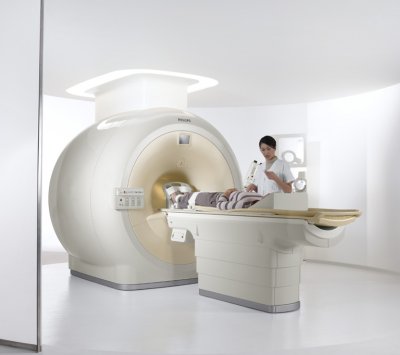
- You will be positioned on a padded table and slowly moved into an open magnet that surrounds the body with a magnetic field.
- Once you are comfortably positioned, it is important that you remain relaxed and completely still during the scan. Movement will result in unclear images.
- There will be a faint knocking, intermittent humming and thumping sounds. These represent changes in the magnetic fields. Earplugs will be provided.
- Breathe normally, as there is nothing about the procedure to make you uncomfortable or painful. You may notice a warm feeling in the area under examination; this is normal but if it bothers you please let us know.
- In some cases, the doctor may order an image enhancement agent. This is a fluid which is injected into a vein probably in your arm. If this is required, it does not mean that your condition is more serious. So do not be concerned.
- You will have voice contact with the radiographer at all times and you can be seen clearly from the control room.
How long will it take?
Depending on the information your doctor needs, the examination can take between 30 to 90 minutes.
What are the benefits and risks?
BENEFITS
- Images of the soft-tissue structures of the body – such as the heart, lungs, liver and other organs – are clearer and more detailed than with other imaging methods.
- MRI can help physicians evaluate the function as well as the structure of many organs.
- The details makes MRI an invaluable tool in early diagnosis and evaluation of tumours.
- MRI contrast material is less likely to produce an allergic reaction than the iodine-based materials used for conventional x-rays and CT scanning.
- MRI enables the detection of abnormalities that might be obscured by bone with other imaging methods.
- MRI provides a fast, non-invasive alternative to x-ray angiography for diagnosing problems of the heart and cardiovascular system.
- Exposure to radiation is avoided.
RISKS
- An undetected metal implant may be affected by the strong magnetic field.
- MRI is generally avoided in the first 12 weeks of pregnancy. Doctors usually use other methods of imaging, such as ultrasound, on pregnant women unless there is a strong medical reason to use MRI.
When you can expect results
The MRI radiologist and radiographer will review the images during the scan to check that they are clear. The report will be sent to your doctor who will then discuss the scan results with you.
Limitations of MRI
- Bone is better imaged by conventional x-rays in some cases and CT is preferred for patients with severe bleeding.
- MRI may not always distinguish between tumour tissue and oedema fluid.
- It does not detect calcium when this is present within a tumour.
- In most cases the examination is safe for patients with metal implants. So patients should inform the staff of the presence of an implant prior to the test.
- The examination must be used cautiously in early pregnancy.
- MRI typically costs more than CT scanning.
Can I use my MediSave?
Yes. Outpatient scans for diagnosis. Claim up to $300 per year per patient.
https://www.cpf.gov.sg/Members/Schemes/schemes/healthcare/medisave

