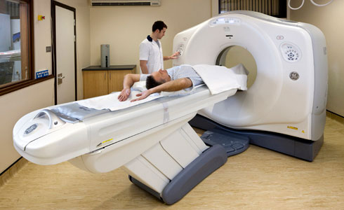
The CT scanner is a specially designed X-ray equipment that rotates around a patient’s anatomy under study. It uses computers which run sophisticated processing algorithms to produce images of the body. An X-ray tube inside the CT scanner rotates on a single axis around the body, emitting an X-ray beam. This beam which passes through the body will be captured by detectors. This beam which passes through the body will be captured by detectors. These detectors are spinning in tandem with the X-ray tube but situated directly opposite to the direction of the X-ray beam.

The radiation beam that passes through the body is attenuated differently, based in the density of the structures. The varying strengths/values received by the detectors will be depicted as varying densities (shades of grey) of the organs that make up the body. The data acquired are usually reconstructed into cross-sectional images (axial plane) of the area being studied. Image manipulation can be performed to show the region being scanned in the sagittal, coronal and/or any oblique planes as required. Special processing algorithms can be applied to better show structures like muscle/soft tissue, blood vessels, bone or lungs. The acquired images may also be processed as a 3-dimensional image which can be manipulated to be viewed from different angles.
The CT scanner provides images of high spatial resolution. Images of different body structures are well differentiated and defined, making CT particularly useful and widely used as an imaging method to find an occult or hidden fracture or provide more information regarding the damage to the bone and adjacent tissues.
The use of CT scanners helps physicians diagnose medical conditions. It is usually minimally invasive and for some examinations, it is a non-invasive procedure. The CT scan can also be used by doctors for interventional procedures to treat medical conditions more accurately.
